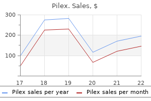
Generic pilex 60 caps lineIn decompensated left-sided coronary heart failure mens health on ipad order 60 caps pilex overnight delivery, the patient might appear dusky (decreased cardiac output) and diaphoretic (sweating because of increased sympathetic nervous activity) prostate cancer journal articles buy 60 caps pilex overnight delivery, and the extremities are cool due to peripheral arterial vasoconstriction. The sample of Cheyne-Stokes respiration may also be current in superior heart failure, characterized by periods of hyperventilation separated by intervals of apnea (absent breathing). The affected person with structural coronary heart disease associated with heart failure however has not but developed signs the patient with current or prior signs of coronary heart failure associated with structural coronary heart illness the affected person with structural heart illness and refractory coronary heart failure symptoms despite maximal medical remedy who requires superior interventions. Sinus tachycardia (resulting from increased sympathetic nervous system activity) can additionally be frequent. In left-sided heart failure, the auscultatory finding of pulmonary raZes ("crackles") is created by the "popping open" of small airways throughout inspiration that had been closed off by edema fluid. This finding is initially apparent at the lung bases, where hydrostatic forces are biggest; however, more severe pulmonary congestion is associated with further rales higher within the lung fields. Compression of conduction airways by pulmonary congestion could produce coarse rhonchi and wheezing; the latter discovering in coronary heart failure is termed cardiac bronchial asthma. Because elevated left heart filling pressures lead to increased pulmonary vascular pressures, the pulmonic part of the second coronary heart sound is often louder than regular. The elevated systemic venous stress produced by proper heart failure is manifested by distention of the jugular veins in addition to hepatic enlargement with abdominal right upper quadrant tenderness. Edema accumulates in the dependent parts of the physique, beginning in the ankles and feet of ambulatory patients and within the presacral areas of those that are bedridden. Pleural effusions could develop in either left- or right-sided heart failure, because the pleural veins drain into each the systemic and pulmonary venous beds. The presence of pleural effusions is recommended on bodily examination by dullness to percussion over the posterior lung bases. This is defined as follows: when a patient is in the upright place, blood move is generally higher to the lung bases than to the apices because of the effect of gravity. Redistribution of circulate happens with the development of interstitial and perivascular edema, as a result of such edema is most prominent on the lung bases (where the hydrostatic strain is the highest), such that the blood vessels within the bases are compressed, whereas move into the higher lung zones is less affected. Depending on the trigger of coronary heart failure, the chest radiograph may present cardiomegaly, defined as a cardiothoracic ratio greater than zero. A excessive proper atrial strain also causes enlargement of the azygous vein silhouette. The reason for heart failure is commonly evident from the historical past, similar to a affected person who has sustained a big myocardial infarction, or by bodily examination, as in a affected person with a murmur of valvular coronary heart illness. Of the several noninvasive checks that may help make this willpower, echocardiography is very helpful and readily available. The 5-year mortality price following the diagnosis ranges between 45% and 60%, with males having worse outcomes than girls. The greatest mortality is due to refractory coronary heart failure, but many patients die abruptly, presumably due to related ventricular arrhythmias. Ventricular dysfunction often begins with an inciting insult, but is a progressive course of, contributed to by the maladaptive activation of neurohormones, cytokines, and steady ventricular transforming. In some patients, this will require surgical repair or replacement of dysfunctional cardiac valves, coronary artery revascularization, aggressive therapy of hypertension, or cessation of alcohol consumption. Elimination of the acute precipitating explanation for signs in a affected person with coronary heart failure who was previously in a compensated state. This could embody, for instance, treating acute infections or arrhythmias, eradicating sources of extreme salt intake, or eliminating medicine that can irritate symptomatology. This is most readily accomplished by dietary sodium restriction and diuretic medications. Measures to increase forward cardiac output and perfusion of significant organs by way of the use of vasodilators and constructive inotropic medication. There is powerful evidence from scientific trials that longevity is enhanced by specific therapies, as described beneath. Diuretics the mechanisms of action of diuretic drugs are summarized in Chapter 17. By promoting the elimination of sodium and water by way of the kidney, diuretics cut back intravascular volume and thus venous return to the heart. Pointe represents the potential additional benefit of mixing an inotrope and vasodilator together. The most essential in coronary heart failure patients embrace overly vigorous diuresis resulting in a fall in cardiac output and electrolyte disturbances (particularly hypokalemia and hypomagnesemia), which can contribute to arrhythmias. In patients with acute heart failure exacerbations, diuretics must be administered intravenously (either by bolus injections or continuous infusion) as a result of venous congestion can limit the absorption of oral diuretics from the gut. As indicated earlier, neurohormonal compensatory mechanisms in coronary heart failure often lead to extreme vasoconstriction, quantity retention, and ventricular remodeling, with progressive deterioration of cardiac function. Moreover, a quantity of studies have proven that sure vasodilator regimens considerably extend survival in patients with coronary heart failure. However, venous vasodilatation in a affected person who is working on the steeper a half of the curve could lead to an undesired fall in stroke quantity, cardiac output, and blood stress. As resistance is reduced by arteriolar vasodilatation, a concurrent rise in cardiac output usually occurs, such that blood stress stays constant or decreases solely mildly. Some groups of medication end in vasodilatation of both the venous and arteriolar circuits ("balanced" vasodilators). Of these, crucial are agents that inhibit the reninangiotensin-aldosterone system. Chronic therapy using the combination of the venous dilator isosorbide dinitrate plus the arteriolar dilator hydralazine has additionally been shown to improve survival in patients with reasonable signs of coronary heart failure. It causes fast and potent vasodilatation, reduces elevated intracardiac pressures, and augments ahead cardiac output. Positive Inotropic Drugs Inotropic medication embrace j3-adrenergic agonists, digitalis glycosides, and phosphodiesterase sort 3 inhibitors (see Chapter 17). Their long-term use is proscribed by the lack of an oral type of administration and by the development of drug tolerance. The latter refers to the progressive decline in effectiveness throughout continued administration of the drug, probably owing to down-regulation of myocardial adrenergic receptors. Despite the initial promise of effective oral phosphodiesterase 3 inhibitors, research so far really reveal lowered survival amongst patients receiving this type of treatment. One of the oldest types of inotropic therapy is digitalis (see Chapter 17), which can be administered intravenously or orally. Digitalis preparations improve contractility, reduce cardiac enlargement, improve signs, and augment cardiac output in sufferers with systolic heart failure. Digitalis also will increase the sensitivity of the baroreceptors, so that the compensatory sympathetic drive in heart failure is blunted, a desired effect that reduces left ventricular afterload. Although digitalis can enhance symptomatology and cut back the rate of hospitalizations in heart failure sufferers, it has not been proven to enhance long-term survival. Thus, its use is proscribed to patients who remain symptomatic regardless of other commonplace therapies or to help slow the ventricular price if atrial fibrillation can be current. The three fl-blockers which have been shown to be useful in randomized scientific trials of coronary heart failure embrace carvedilol (a nonselective ~-blocker with weak a-blocking propertiessee Chapter 17) and the ~1-selective brokers metoprolol succinate and bisoprolol. Nonetheless, fl-blockers should all the time be used cautiously in heart failure to forestall acute deterioration associated to their adverse inotropic effect. Aldosterone Antagonist Therapy There is evidence that chronic extra of aldosterone in heart failure contributes to cardiac fibrosis and opposed ventricular remodeling.
Buy pilex 60 caps low costIn dilatation of the basis of the aorta or pulmonary artery prostate cancer youtube pilex 60 caps buy, the sound is associated with sudden tensing of the aortic or pulmonic root with the onset of blood move into the vessel prostate ultrasound and biopsy pilex 60 caps lowest price. In distinction, the pulmonic ejection click is heard only at the base, and its intensity diminishes throughout inspiration (see Chapter 16). An ejection click follows the opening of the aortic or pulmonic valve in cases of valve stenosis or dilatation of the corresponding nice artery. Opening Snap Opening of the mitral and tricuspid valves is generally silent, but mitral or tricuspid valvular stenosis (usually the outcome of rheumatic heart illness; see Chapter 8) produces a sound, termed as snap, when the affected valve opens. On inspiration, regular splitting of the second heart sound (52) is noticed in order that three sounds are heard. Because of its proximity to A2, the A2-0S sequence could be confused with a broadly break up second coronary heart sound. This occurs as a result of the diploma of left atrial stress elevation corresponds to the severity of mitral stenosis. When the ventricle relaxes in diastole, the greater the left atrial strain, the earlier the mitral valve opens. Compared with extreme stenosis, gentle illness is marked by a less elevated left atrial pressure, lengthening the time it takes for the left ventricular strain to fall below that of the atrium. A left-sided S3 is often loudest over the cardiac apex while the patient lies in the left lateral decubitus position. Production of the 53 appears to result from tensing of the chordae tendineae during fast filling and enlargement of the ventricle. In these groups, an S3 implies the presence of a supple ventricle able to normal fast growth in early diastole. Conversely, when heard in middle-aged or older adults, an S3 is an indication of disease ensuing from a dilated ventricle e. This sound is generated by the left (or right) atrium ejecting blood into a stiffened ventricle. Thus, an 54 often signifies the presence of cardiac disease-specifically, a lower in ventricular compliance typically ensuing from ventricular hypertrophy or myocardial ischemia. Like an S3, the S4 is a uninteresting, low-pitched sound and is finest heard with the bell of the stethoscope. In the case of the extra frequent left-sided S4, the sound is loudest on the apex, with the patient lying in the left lateral decubitus position. Quadruple Rhythm or Summation Gallop In a affected person with both an fifty three and S4, these sounds, in conjunction with S1 and eighty two, produce a quadruple beat. If a patient with a quadruple rhythm develops tachycardia, diastole turns into shorter in period, the S3 and S4 coalesce, and a summation gallop outcomes. The summation of 53 and 54 is heard as a protracted middiastolic, low-pitched sound, typically louder than S1 and Sz. The Cardiac Cycle: Mechanisms of Heart Sounds and Murmurs 35 Pericardial Knock A pericardia! It outcomes from the abrupt cessation of ventricular filling that occurs when the increasing ventricle meets a inflexible pericardium in early diastole, which is the hallmark of constrictive pericarditis. Under normal situations, the motion of blood by way of the vascular mattress is laminar, smooth, and silent. However, on account of hemodynamic and/or structural modifications, laminar move can become disturbed and produce an audible noise. Abnormal shunting of blood from one vascular chamber to a lower-pressure chamber. Timing refers to whether or not the murmur occurs throughout systole or diastole, or is steady. High-frequency murmurs are brought on by giant stress gradients between chambers. For example, a crescendo-decrescendo (or "diamond-shaped") murmur first rises after which falls off in intensity. Examples of the consequences of maneuvers on particular murmurs are presented in Chapter 8. It begins after the first heart sound and terminates before or during fifty two, depending on its severity and whether or not the obstruction is of the aortic or pulmonic valve. Ejection murmurs are crescendodecrescendo in configuration (A), whereas pansystolic murmurs are uniform all through systole (B). A Late systolic murmur often follows a midsystolic click on and suggests mitral (or tricuspid) valve prolapse (C). This gap corresponds to the period of isovolumetric contraction of the left ventricle Aorta (the interval after the mitral valve has closed however before the aortic valve has opened). The murmur becomes extra intense as flow increases throughout the aortic valve during the rise in left ventricular strain (crescendo). Systolic ejection murmur of aortic reflecting the sizable pressure gradient across the stenosis. It is greatest heard in the "aortic area" at the second heart sound (51) and the onset of the munnur (first and third proper intercostal areas close to the sternum dashed line). The murmur sometimes radiates towards the neck (the course of turbulent blood flow) but typically could be heard in a large distribution, together with the cardiac apex. The murmur of pulmonic stenosis also begins after 51 � It could also be preceded by an ejection click, however not like aortic stenosis, it may lengthen past the A2 sound. Pulmonic stenosis is normally loudest on the second to third left intercostal areas near the sternum. Young adults usually have benign systolic ejection murmurs also termed "harmless murmurs") resulting from elevated systolic circulate throughout regular aortic and pulmonic valves. This kind of murmur typically turns into softer or disappears when the affected person sits upright. Pansystolic (also termed holosystolic) murmurs are attributable to regurgitation of blood throughout an incompetent mitral or tricuspid valve or via a ventricular septal defect (see Chapter 16). In mitral and tricuspid valve regurgitation, as quickly as ventricular systolic pressure exceeds atrial stress. The pansystolic murmur of superior mitral regurgitation continues by way of the aortic closure sound as a outcome of left ventricular stress remains larger than that in the left atrium on the time of aortic closure. It typically radiates to the best of the sternum and is excessive pitched and blowing in high quality. The depth of the murmur increases with inspiration as a outcome of the negative intrathoracic strain induced during inspiration enhances venous return to the guts. The latter augments right ventricular stroke volume, thereby growing the quantity of regurgitated blood. The most typical instance is mitral regurgitation brought on by mitral valve prolapse-bowing of abnormally redundant and elongated valve leaflets into the left atrium during ventricular contraction. This murmur is often preceded by a midsystolic click on and is described in Chapter 8. The severity of aortic stenosis impacts the shape of the systolic murmur and the heart sounds. Early diastolic murmurs end result from regurgitant flow through either the aortic or pulmonic valve, with the former being far more common in adults. If produced by cwrtic valve regurgitation, the murmur begins at A2, has a decrescendo shape, and terminates before the following S1� Because diastolic relaxation of the left ventricle is speedy, a pressure gradient develops instantly between the aorta and lower-pressured left ventricle in patients with aortic regurgitation, and the murmur due to this fact shows its maximum intensity at its onset.
Diseases - Whitaker syndrome
- Davis Lafer syndrome
- Hyde Forster Mccarthy Berry syndrome
- Cilliers Beighton syndrome
- Female sexual arousal disorder
- Edwards syndrome
60 caps pilex cheap otcManagement of extragonadal germ cell tumors and the importance of bilateral testicular biopsies prostate cancer uk discount 60 caps pilex mastercard. Management of seminomatous testicular cancer: a binational potential populationbased study from the Swedish norwegian testicular cancer research group prostate 180 at walgreens cheap pilex 60 caps with amex. Testicular carcinoma in situ in sufferers with extragonadal germ-cell tumours: the clinical position of pre-treatment biopsy. Optimal planning goal quantity for stage I testicular seminoma: a Medical Research Council randomized trial. The International Germ Cell Consensus Classification: a prognostic issue based mostly staging system for metastatic germ cell cancer. Management of postchemotherapy residual mass in sufferers with advanced seminoma: Indiana University expertise. Integrated approach to the management of sufferers with advanced germ cell tumors of the testis. Management of residual mass in superior seminoma: results and recommendations from the Memorial Sloan Kettering Cancer Center. Is post chemotherapy resection of seminomatous components related to greater acute morbidity Estimating the chance of most cancers related to imaging associated radiation throughout surveillance for stage I testicular most cancers utilizing computerized tomography. Recent stories on the impact of low doses of ionizing radiation and its dose-effect relationship. A novel surveillance protocol for stage I nonseminomatous germ cell testicular tumours. Medical Research Council prospective examine of surveillance for stage I testicular teratoma. Prognostic elements that determine patients with medical stage I nonseminomatous germ cell tumors at low danger and excessive danger for metastasis. Randomized trials in 2466 patients with stage I seminoma: patterns of relapse and follow-up. Pelvic recurrence in stage I seminoma: a model new phenomenon that questions trendy protocols for radiotherapy and follow-up. The results of radiotherapy treatment adjustments on sites of relapse in stage I testicular seminoma. Second cancers amongst forty,576 testicular most cancers sufferers: give consideration to long-term survivors. Treatmentspecific dangers of second malignancies and heart problems in 5-year survivors of testicular cancer. Risk-adapted management for sufferers with scientific stage I seminoma: the Second Spanish Germ Cell Cancer Cooperative Group examine. Posttreatment surveillance after paraaortic radiotherapy for stage I seminoma: a systematic evaluation. Radiotherapy versus singleagent arboplatin in adjuvant therapy of stage I seminoma: a randomised trial. Risk factors for relapse in clinical stage I nonseminomatous testicular germ cell tumors: results of the German Testicular Cancer Study Group Trial. Prospective metastatic risk assignment in scientific stage I nonseminomatous germ cell testis most cancers: a single establishment pilot study. Non-risk-adapted surveillance for sufferers with stage I nonseminomatous testicular germ-cell tumors: diminishing treatment-related morbidity while sustaining efficacy. Outcomes of surveillance protocol of medical stage I nonseminomatous germ cell tumors-is shift to threat tailored coverage justified Minimizing remedy with out compromising remedy with main surveillance for scientific stage I embryonal predominant carcinoma noneminomatous testicular cancer: a inhabitants primarily based analysis from British Columbia. Treating stage I nonseminomatous germ cell tumours with a single cycle of chemotherapy. Long-term followup outcomes of 1 cycle of adjuvant bleomycin, etoposide and cisplatin chemotherapy for prime risk clinical stage I nonseminomatous germ cell tumors of the testis. Retroperitoneal lymph node dissection for nonseminomatous germ cell testicular most cancers: impact of patient choice elements on end result. Retroperitoneal lymph node dissection within the remedy of low-stage nonseminomatous germ cell tumors of the testicle: an update. Retroperitoneal lymphadenectomy for testis tumor with nerve sparing for ejaculation. Primary retroperitoneal lymph node dissection in scientific stage A nonseminomatous germ cell eighty one. Retroperitoneal lymph node dissection in patients with low stage testicular most cancers with embryonalcarcinoma predominance and/or lymphovascular invasion. Retroperitoneal lymph node dissection with no adjuvant chemotherapy in clinical stage I nonseminomatous germ cell tumours: Long-term outcome and evaluation of risk factors of recurrence. Laparoscopicretroperitoneal lymph node dissection: does it nonetheless have a task in the administration of scientific stage I nonseminomatous testis cancer Complications of primary nerve sparing retroperitoneal lymph node dissection for clinical stage I nonseminomatous germ cell tumors of the testis: expertise of the German Testicular Cancer Study Group. Failure of high-dose cyclophosphamide and etoposide combined with double-dose cisplatin and bone marrow help in patients with high-volume metastatic nonseminomatous germ-cell tumours: mature results of a randomised trial. High-dose carboplatin, etoposide, and cyclophosphamide with autologous bone marrow transplantation in first-line therapy for patients with poor-risk germ cell tumors. Post-chemotherapy lymph node histology in radiologically normal patients with metastatic nonseminomatous testicular cancer. Adjunctive surgical procedure after chemotherapy for nonseminomatous germ cell tumors: recommendations for patient selection. Resection of postchemotherapy residual masses and limited retroperitoneal lymphadenectomy in sufferers with metastatic testicular nonseminomatous germ cell tumors. Does necrosis on frozen-section analysis of a mass after chemotherapy justify a restricted retroperitoneal resection in sufferers with advanced testis most cancers Distribution of nodal metastases after chemotherapy in nonseminomatous testis cancer: a potential indication for restricted dissection. Pathologic findings and clinical consequence of patients present process retroperitoneal lymph node dissection after multiple chemotherapy regimes for metastatic testicular germ cell tumors. Is full bilateral retroperitoneal lymph node dissection all the time essential for postchemotherapy residual tumor Postchemotherapy retroperitoneal lymph node dissection in advanced testicular most cancers: radical or modified template resection. Retroperitoneal recurrences after retroperitoneal lymph node dissection for low-stage nonseminomatous germ cell tumors. Reoperative retroperitoneal surgical procedure for nonseminomatous germ cell tumor: medical presentation, patterns of recurrence and outcome. Outcome analysis for patients with elevated serum tumor markers at postchemotherapy retroperitoneal lymph node dissection. En bloc nephrectomy in sufferers present process post-chemotherapy retroperitoneal lymph node dissection for nonseminomatous testis most cancers: indications, implications and outcomes.
Purchase pilex 60 caps fast deliveryUltrasound can additionally be frequently used to characterize incidental lesions discovered on other imaging modalities as strong or cystic man health news discount pilex 60 caps. Technique and regular appearances To achieve correct diagnosis requires consideration to good ultrasound method mens health arm workout 60 caps pilex cheap overnight delivery. Echopoor medullary pyramids (*) and echogenic renal sinus fat (s) may be appreciated. Congenital abnormalities and pseudomasses Numerous congenital abnormalities and pseudomasses of the kidneys have been described and can be demonstrated with ultrasound. Pelvic kidneys are found in between 1 in 2,200 and 1 in 3,000 sufferers, are more frequent on the left facet and are prone to ureteropelvic junction obstruction and formation of calculi. The most typical fusion abnormality is the horseshoe kidney, which occurs in 1 in four hundred people. Crossed fused renal ectopia is sometimes additionally seen on ultrasound as an enlarged elongated unilateral kidney with two distinct renal sinus complexes, typically with a different orientation. Duplex kidneys are one of the more frequent congenital abnormalities present in between 2. The higher moiety could additionally be hydronephrotic due to obstruction by a ureterocoele,14 which ought to be sought on bladder scanning. The lower moiety could have a dilated collecting system or cortical scarring secondary to reflux. Hypertrophied columns of Bertin represent a protrusion of regular parenchymal tissue into the renal sinus and are a regular discovering on ultrasound, seen in almost half of kidneys in a single examine. Splenic (Dromedary) humps are a focal bulge of regular renal parenchyma arising from the interpolar area of the kidney, often on the left facet. Many patients also present common indentations of the renal contour representing persistent foetal lobulation. These situations have to be recognized as a traditional variant rather than a renal mass or scarring. Ureteric obstruction and renal stone illness Ultrasound is incessantly requested to rule out obstruction, either as a cause of renal useful impairment or in sufferers with flank pain. Ultrasound is extremely correct in detecting hydronephrosis,18 but it ought to be remembered that ultrasound demonstrates anatomical quite than functional modifications. There is also a delay in the restoration of regular appearances after aid of an obstruction, typically for several days, and a level of pelvicalyceal dilatation could persist indefinitely, notably after reduction of longstanding or extreme obstruction. A variety of grading methods have been proposed for the severity of hydronephrosis however simply stating gentle, reasonable, or extreme is usually adequate. The accuracy of ultrasound in diagnosing renal obstruction could be improved by the use of Doppler strategies. Elevated intrarenal vascular resistance in an obstructed kidney can be demonstrated by spectral Doppler examination. Within the kidney it could be troublesome to distinguish a small stone from the echogenic renal sinus. Sensitivity is improved through the use of the next frequency transducer and making certain the main focus is situated at the degree of the suspected calculus. Ultrasound has a reported sensitivity of 96% and specificity of 89% for detection of stones within the pelvicalyceal system (with tomography as the gold standard),27 however the determine is as little as 37% for ureteric calculi,28 largely because of overlying bowel fuel. In feminine patients transvaginal ultrasound can be utilized to demonstrate distal ureteric stones. They may be parapelvic, intraparenchymal or exophytic, in which case observing motion with the kidney on respiration is helpful in making the prognosis. Note the multiple thickened and enhancing septa (examples arrowed) within the cyst raising issues for a cystic malignancy. Benign cysts should be distinguished from cystic malignancy; septations, loculations, or solid elements enhance the probability of malignancy. A variety of different non-malignant renal lesions could appear as complex cystic plenty on ultrasound together with multilocular cystic nephroma, hydatid disease, abscess, haematoma, and xanthogranulomatous pyelonephritis. Smaller tumours may be hyperechoic and impossible to distinguish from angiomyolipomas. Other stable renal lots include angiomyolipomata, which are usually brightly echogenic on ultrasound because of the presence of fats. Oncocytomas are often inconceivable to distinguish from renal cell carcinoma on ultrasound, although a central scar is said to be characteristic and colour Doppler ultrasonography may present central radiating vessels. The primary function of ultrasound in this setting is to measure renal size, assess parenchymal thickness, exclude obstruction and, when applicable, to guide biopsy. The findings might embody decreased parenchymal echogenicity and increase in renal measurement. Perfusion defects could additionally be detected using color or power Doppler; nevertheless the scan is regularly regular. In focal pyelonephritis (lobar nephronia) a part of the kidney could additionally be enlarged or of altered echogenicity, either elevated, mixed, or decreased. When the perinephric soft tissues are involved the whole kidney could additionally be tough to determine. Chronic pyelonephritis secondary to vesico-ureteric reflux may cause renal scarring, seen on ultrasound as focal cortical thinning overlying a dilated or clubbed calyx. Ultrasound is important in detection of the issues of renal transplantation, and is used both for regular surveillance and as the first line imaging investigation for graft dysfunction. Renal transplants are sometimes positioned in both iliac fossa, but the axis of the kidney is variable and the scan plane should be tailor-made to accommodate this. The superficial position of the transplant often allows it to be simply assessed and a better frequency transducer can often be used. The scan method includes an intensive greyscale evaluation of the transplant and analysis of the encircling soft tissues for fluid collections. Global renal perfusion is then assessed with colour Doppler adopted by a spectral Doppler evaluation of a number of interlobar arteries. In the early postoperative interval peritransplant collections might symbolize urinoma, haematoma, lymphocoele, or abscess formation; ultrasound guided needle aspiration may be necessary to differentiate between them. Renal artery thrombosis (global or segmental) is definitely recognized by an absence of perfusion of all or part of the transplant. Arteriovenous fistulae and pseudoaneurysms are often related to biopsy and are readily recognized with Doppler methods. The key findings are lack of continuity of the renal cortex, perinephric collections of blood or urine, and disruption of renal perfusion. Medical renal disease the various causes of medical renal illness have similar sonographic appearances- enlarged or normal measurement kidneys with increased echogenicity of the cortex. Peak systolic circulate velocities of >250 cm/s in the main transplant artery indicate a haemodynamically significant stenosis.
Buy generic pilex 60 capsThe amount of power deposited divided by the mass of tissue uncovered known as the absorbed dose mens health events cheap 60 caps pilex. The unit of absorbed dose is joule per kilogram (J/kg) however for convenience it has been given a particular name: grey [Gy] prostate cancer fund buy pilex 60 caps line. Equivalent dose Different kinds of radiation cause different biological effects for the same amount of vitality deposited. For instance neutrons or alpha particles cause extra damage per unit absorbed dose in comparability with X-rays or gamma rays. To permit for this a quality factor for radiation was introduced resulting within the equivalent dose. The unit of equal dose is also joule per kilogram (J/kg) however to keep away from confusion it has been given a special name: sievert [Sv]. Effective dose In addition tissues have completely different sensitivity to radiation as a outcome of cell turnover fee and tissue particular characteristics. To account for this a tissue weighting factor was added to modify equal dose ensuing in the efficient dose. Doses in medical imaging and radiation safety are normally organ particular and check with effective doses. Definition of ionizing radiation Radiation is a form of energy transmitted via house. There are many forms of radiation corresponding to warmth, mild, radio waves, microwaves, X-rays, and gamma rays. The frequency of the waves describes its place in the electromagnetic spectrum. Low vitality radio waves with low frequencies are at one finish of the electromagnetic spectrum; high frequency, high power waves such as X-rays or gamma rays at the other finish. High frequency waves that carry a appreciable quantity of vitality can penetrate materials and transfer energy to an atom that may cause displacement of an electron from its orbit across the nucleus. The probability of a tissue or organ to undergo results from ionizing radiation is called radiosensitivity. Tissues with high charges of mitosis and undifferentiated cells are essentially the most sensitive. Therefore bone marrow cells, gonads, lens cells, and bowel mucosal cells are extremely sensitive whereas bone and neural cells are comparatively much less radiosensitive. Natural background radiation Natural background radiation is throughout us and includes excessive vitality cosmic rays, radioactive nuclides from the earth crust such as uranium and thorium and radon gasoline. The annual effective dose from pure background radiation varies substantially all through the nation and the world as a outcome of variation in altitude and the constituents of the soil. The average annual efficient dose from natural sources on the planet is estimated to be 2. Tissue reactions (deterministic effects) Ionizing radiation may cause acute results that are largely as a result of cell death. These are also referred to as deterministic or non-stochastic results and are often seen at excessive radiation doses such radiotherapy or radiation accidents. Because a threshold could be identified, radiation protection measures and occupational dose limits can get rid of such effects for workers. Deterministic effects embrace pores and skin erythema, ulceration and burns, bone marrow depression, cataracts, acute radiation illness, sterility, and foetal dying. However the largest amount of artificial radiation publicity is through medical imaging procedures. Those effects are random and long-term, and also referred to as stochastic or non-deterministic effects. The first examine investigating the direct danger of most cancers induction from medical imaging was printed in 2012. Medical radiation procedures have been 65 instances extra frequent in developed international locations in comparison with underdeveloped international locations reflecting an imbalance of radiation exposure and healthcare provision. In several countries medical radiation publicity is now bigger than publicity from pure sources. Extraordinarily giant numbers of sufferers would need to be adopted up over a lifetime to quantify threat at low doses of radiation exposure. The assumption that radiation risk continues in a linear style when extrapolated from high to low doses is termed the linear non-threshold model. It assumes that no level of radiation publicity is protected and that even the smallest dose of radiation has a possible to trigger harm. Potential well being results of ionizing radiation Biological effects As previously described high energy ionizing radiation causes displacement of an electron from its orbit across the nucleus. Single strand breaks could simply be repaired with out detrimental effects to the cell. In a population, radiation results particularly from low dose radiation can be obscured by the large variety of nonradiation cancers occurring: 1 in three folks will develop cancer from pure causes and 1 in 6 people are predicted to die of such most cancers. The mean latent interval for leukaemia is 7�10 years, for bone tumours 10�15 years, and approximately 20 years for many stable tumours. However radiationinduced mutations can theoretically happen in reproductive cells (egg and sperm) resulting in hereditable diseases. Concerns about radiation and induction of most cancers are more doubtless to be in the public focus for the foreseeable future. It is due to this fact the duty of all healthcare professionals to handle and management the chance to shield patients. Radiation exposure very early on in a pre-implantation stage might lead to an all or nothing phenomenon of spontaneous abortion or normal pregnancy. Malformations are thought to have a threshold of 100�200 mGy absorbed dose which is never reached in medical diagnostic imaging. The Ionising Radiation (Medical Exposure) Regulations 2000 the Ionising Radiation (Medical Exposure) Regulations 2000 impose obligations on hospital authorities and clinicians to minimize radiation doses to patients. However, considering the advantages from imaging in diagnosing illness and damage, a medically indicated imaging research will nearly all the time outweigh the chance associated with it. This is particularly apparent when evaluating risks related to frequent day by day activities such as driving a automobile. These ranges point out the radiation dose a typical affected person is prone to obtain from a normal radiological examination. Radiation doses from interventional procedures can be high, significantly to the skin the place the same space is persistently irradiated during an prolonged procedure. Sometimes it can be beneficial simply to observe the affected person for a time interval for symptoms to unmask. The iRefer: Making the most effective use of medical radiology publication by the Royal College of Radiologists might help clinicians to choose applicable imaging. Minimizing danger to workers Principles Any measures that reduce affected person dose additionally scale back potential occupational dose to employees. The inverse square regulation demonstrates that doubling the distance to the source reduces the dose by a factor of four.
60 caps pilex cheap with mastercardNeurologic unwanted side effects embody proximal muscle weak spot man health 1st 60 caps pilex free shipping, peripheral neuropathy mens health xbox game pilex 60 caps discount free shipping, ataxia, tremors, and sleep disturbances. Corneal microdeposits may be detected in sufferers receiving chronic amiodarone therapy, however these hardly ever affect vision. Amiodarone interacts with, and increases the activity of, sure medicine together with warfarin and digoxin, such that the dosages of those agents must be adjusted. Similar to amiodarone, it blocks potassium, sodium, and L-type calcium channels and inhibits p- and a-adrenergic receptors. It is run orally and reaches a gentle state in four to 8 days, a lot quicker than oral amiodarone. The main unwanted facet effects are gastrointestinal, together with nausea, vomiting, and diarrhea. It prolongs the duration of the action potential, will increase the refractory period of atrial and ventricular tissue, and inhibits conduction in accent bypass tracts. It is efficient in the treatment of both supraventricular and ventricular arrhythmias. Because sotalol is excreted exclusively by the kidneys, its dosage ought to be adjusted in the presence of renal disease. This complication happens in roughly 2% of sufferers and is more frequent in patients with a historical past of heart failure and in ladies for unknown reasons). Dofetilide is excreted by the kidney, and its dose must be adjusted in sufferers with renal failure. This agent prolongs the motion potential duration and will increase atrial and ventricular refractoriness. At one time, intravenous verapamil was the remedy of alternative for acute episodes of such rhythms, but intravenous adenosine (described within the next section) has assumed that function. The most necessary side effect of verapamil and diltiazem, when administered intravenously, is hypotension. In addition, these brokers ought to be avoided, or used cautiously, in patients receiving! J-blocker remedy, because the combined unfavorable inotropic and chronotropic results might precipitate coronary heart failure and/or significant bradycardia. Adenosine additionally inhibits membrane adenylate cyclase activity, by way of the G protein a,-subunit. Conversely, dipyridamole interferes with mobile uptake and degradation of adenosine and therefore amplifies its impact. With a half-life of only 10 seconds, adenosine has very transient unwanted facet effects (headache, chest ache, flushing, bronchoconstriction). Because methylxanthines (caffeine, theophylline) competitively antagonize the adenosine receptor, greater doses of adenosine may be essential in sufferers using these substances. Conversely, dipyridamole inhibits the breakdown of adenosine and amplifies its effect. In coronary heart failure, enhanced renal reabsorption of sodium and water, with subsequent expansion of the exttacellular quantity, contributes to peripheral edema and pulmonary congestion. In the treatment of hypertension, diuretics equally reduce intravascular volume and in some circumstances promote vascular dilatation. In the kidney, the rate of glomerular filtration usually averages one hundred thirty five to one hundred eighty L/day in normal adults. Approximately 65% to 70% of the filtered Na+ is reabsorbed isosmotically within the proximal tubule by lively transport. Approximately 70% of filtered sodium is reabsorbed within the proximal convoluted tubule, 25% within the thick ascending limb of the loop of Henle, S% within the distal convoluted tubule, and 1% to 2% within the cortical collecting tubule (mediated by the motion of aldosterone). Diuretic medicine are secreted into the proximal convoluted tubule and act on the websites proven. In the distal convoluted tubule, an extra small fraction of NaCl is reabsorbed (approximately 5%). In the cortical collecting duct, Na+ permeability is modulated by an aldosterone-sensitive mechanism, such that Na+ is reabsorbed into the tubular cells in the presence of aldosterone, creating a lumen-negative potential distinction that enhances K+ and H+ excretion. In the amassing tubule, nevertheless, water permeability and reabsorption are promoted by antidiuretic hormone and driven by the osmotic gradient between the tubule and the hypertonic interstitium. Therefore, substances that intrude with antidiuretic hormone, such as ethanol, have diuretic actions. These classes are distinguished by the positioning of the kidney tubule the place they act and by their potency. Loop diuretics impair absorption within the thick ascending limb of the loop of Henle, thiazide diuretics act on the distal tubule and accumulating phase, and potassium-sparing diuretics act on the aldosteronesensitive area of the cortical amassing tubule. Members of a fourth group, the carbonic anhydrase inhibitors, are weak diuretics not often used within the remedy of hypertension or heart failure. They act on the proximal convoluted tubule, leading to a loss of bicarbonate (and sodium) in the urine. Cardiovascular Drugs forty three 7 Loop Diuretics these agents are so named because they act principally on the thick ascending limb of the loop of Henle. They are highly effective diuretics that outcome within the excretion of 20% to 25% of the filtered Na+ load by way of inhibition of the Na+-2cl-K+ cotransport system. Because inhibition at this web site impairs the technology of a hypertonic interstitium, the gradient for passive water motion out of the amassing duct is diminished and water diuresis outcomes. Loop diuretics are of great significance in the acute administration of pulmonary edema (administered intravenously) and in the treatment of persistent heart failure or peripheral edema (taken orally). Unlike different diuretics, they have an inclination to be effective within the setting of impaired renal operate. In addition to the diuretic effect, and even preceding it, drugs of this class may induce venous vasodilation, which can be useful in lowering venous return to the center and pulmonary congestion (see Chapter 9). The mechanism of venous vasodilation seems to contain drug-induced prostaglandin and nitric oxide generation from endothelial cells, which act to chill out vascular easy muscle (see Chapter 6). The most typical unwanted facet effects of the loop diuretics are intravascular volume depletion, hypokalemia, and metabolic alkalosis. Hypokalemia arises as a outcome of (1) these agents impair the reabsorption of sodium in the loop of Henle, such that an elevated quantity of Na+ is delivered to the distal tubule, the place it prompts greater-than-normal exchange for potassium (and therefore more K+ excretion into the urine) and 2) diuretic-induced intravascular volume depletion activates the renin-angiotensin system. Hypomagnesemia may end result, as a outcome of magnesium reabsorption depends on NaCl transport in the thick ascending limb of the loop of Henle, the motion blocked by these drugs. It is believed to come up from electrolyte disturbances of the endolymphatic system, most probably because of Na+-2cl- -K+ cotransport inhibition by the diuretic at that web site. The mostly used loop diuretic is furosemide, the oral type of which demonstrates reliable gastrointestinal absorption however a brief duration of action (4 to 6 hours) that limits its usefulness in the chronic therapy of hypertension. Bumetanide is just like furosemide and shares its actions and antagonistic effects however has larger efficiency and bioavailability. It also appears to have a lower incidence of ototoxicity than the opposite drugs of this class. Ethacrynic acid is the one nonsulfonamide loop diuretic, so it can be prescribed to sufferers with sulfonamide intolerance.
Squaw Weed (Golden Ragwort). Pilex. - How does Golden Ragwort work?
- Dosing considerations for Golden Ragwort.
- What is Golden Ragwort?
- Are there any interactions with medications?
- Diabetes, high blood pressure, spasms, fluid retention, bleeding, irregular or painful menstrual periods, and other uses.
- Are there safety concerns?
Source: http://www.rxlist.com/script/main/art.asp?articlekey=96659
60 caps pilex order with mastercardDiagnostic accuracy of noninvasive exams to consider bladder outlet obstruction in men: detrusor wall thickness man health daily lifestyle category effective 60 caps pilex, uroflowmetry prostate exam procedure 60 caps pilex fast delivery, postvoid residual urine, and prostate quantity. Effect of bladder carcinoma location on detection rates by ultrasonography and computed tomography. Outcome of isolated antenatal hydronephrosis: A systematic evaluate and meta-analysis. Differential prognosis of neonatal adrenal haemorrhage and congenital neuroblastoma by color coded Doppler sonography and power Doppler sonography. It has since advanced to be used to get hold of volumes of picture knowledge of the related body section, which can be manipulated with image processing software program to view the internal organs on sub-millimetre sections in multiple planes, and a whole host of other applications together with surface rendering. The gantry rotates around the patient and X-rays are exposed to acquire information from different angles, which are used to assemble the image using picture reconstruction algorithms. The velocity of acquisition can be boosted by means of multiple rows of detectors, allowing the whole physique to be imaged in a matter of seconds. For instance, to evaluate bone, a window range set around the aforementioned limits would demonstrate bone in detail with all constructions less dense than bone greyed out. There are normally preset window settings for different organs and tissue sorts on imaging workstations, with the operate to fine-tune the vary accordingly. Low dose methods are routinely employed, having been demonstrated to maintain high rates of accuracy for detecting lithiasis regardless of the compromise in image quality of sentimental tissue constructions. The examination requires the administration of intravenous distinction and routinely entails two phases, an unenhanced and nephrographic phase. It permits for baseline characterization of the density of the lesion to establish enhancement patterns (if any) and detection of high density components which can be in any other case obscured with the administration of distinction. The nephrographic section is acquired 100 seconds after administration of intravenous contrast. It produces homogeneous enhancement of the renal parenchyma, permitting for dependable detection of renal plenty. The nephrographic phase is acquired of the kidneys (as previously described) after administration of intravenous distinction, allowing for detection of renal lots. The excretory section of the study assesses the amassing methods, ureters and, to a lesser extent, the urinary bladder when optimum luminal opacification of these structures has taken place following distinction excretion by the kidneys. As with the administration of any drug, patient consent have to be obtained prior to the examination. A described variation of the technique allows acquisition of each the nephrographic and excretory section in a single series by administering two boluses of intravenous contrast after the unenhanced phase. Both doses are administered 10 minutes aside with image acquisition performed after a suitable nephrographic phase delay from the second dose. At this time point, the kidneys can be imaged within the nephrographic part and contrast from the first dose would have begun to bear renal excretion. This affords a radiation dose saving to the affected person and improves workflow by reducing scanning time. Patients should be triaged based on a stringent choice standards, and the place this raises doubt relating to the working prognosis of renal colic, different imaging evaluation ought to be contemplated. This is of specific relevance to younger female sufferers the place another gynaecological cause is prevalent. Published series have revealed that the detection price of lithiasis is mostly higher in males than females (55�61. Contrast brokers could also be classified in accordance with their osmolarity (high or low) and composition (ionic or non-ionic). Both characteristics contribute to the brokers osmolality (and therefore the propensity to cause fluid shifts), the highest being excessive osmolar ionic agents and lowest being low osmolar non-ionic. Essential points to cowl are a historical past of earlier contrast response, bronchial asthma, renal disease, diabetes mellitus, allergy symptoms, and metformin remedy. The alternative of imaging investigation is dictated by the pre-test chance, which is influenced by two major elements: age and nature of haematuria. In all-age cohorts investigated for macroscopic haematuria, the prevalence of sufferers identified with urological cancer and under 40 years has been reported to be zero. The choice of imaging investigation is somewhat much less clear-cut within the context of microscopic haematuria. Apart from persistence of microscopic haematuria, the presence of pain in this context provides more weight, with the prevalence of urolithiasis in sufferers presenting with asymptomatic microscopic haematuria reported to be solely 4�4. It would thus be affordable to carry out an ultrasound within the youthful affected person with microscopic haematuria (to assess kidney measurement, look for hydronephrosis and ancillary indicators of renal parenchymal disease). Percutaneous nephrolithotomy planning the excretory phase offers a transparent road map for the method to percutaneous nephrolithotomy by depicting the position and burden of calculi throughout the pelvicalyceal system, the anatomy of which could be advanced. The incidence of metastatic illness is positively related to tumour measurement with lung being the most common website of unfold. These include biochemical failure after remedy with healing intent and where symptoms elevate the potential for extra-osseous metastatic illness. The thin slice axial images ought to be reviewed first, with close scrutiny of the renal parenchyma and accumulating system for calculi, which would appear as excessive density foci. Both ureters must be traced down to their factors of insertion into the urinary bladder. The urinary bladder should be reviewed for vesical calculi, significantly throughout the dependent a part of the bladder. It also has the advantage of revealing extra-genitourinary findings, which can be of direct relevance to the higher tract abnormality. There are additionally residual renal calculi predominantly within the lower pole calyx (arrow). This is of significance in assessing the maximum dimension of calculi, which may be underestimated on axial images, in flip influencing patient management. Anatomical relationships of areas of pathology may also be better represented on totally different projections, notably coronal photographs, for pre-procedural planning. Review of the opposite stomach organs is essential, notably when no urinary tract abnormalities are detected. This is established by comparing the density of the lesion on the unenhanced section to the density on the nephrographic phase. The density is measured using a region-of-interest device on the imaging workstation to interrogate the interior content material of the lesion. Lymphoma and metastatic illness typically feature as a quantity of bilateral stable lots. When evaluating a cystic renal mass, the well- established Bosniak renal cyst classification is applied. Each class has accompanying scientific management recommendations which ought to by no means be adhered to strictly. The urinary tract should be scrutinized from the higher pole calyx right down to the bladder base. Wide windowing is crucial to avoid lacking small urothelial lesions which might in any other case be obscured by contrast.
Cheap 60 caps pilex with amexDistinction between obstructive and nonobstructive pyelocaliectasis with duplex Doppler sonography prostate oncology 21 60 caps pilex discount. Diagnosis of acute ureteral calculus obstruction in pregnant ladies using colour and pulsed doppler sonography mens health edinburgh 2012 pilex 60 caps purchase on line. Evaluation of painful hydronephrosis in pregnancy: magnetic resonance urographic patterns in physiological dilatation versus calculous obstruction. Testicular microlithiasis: is there a necessity for surveillance within the absence of different danger factors Testicular microlithiasis prevalence and tumor danger in a inhabitants referred for scrotal sonography. Scrotal calcification in a symptomatic paediatric population: prevalence, location, and look in a cohort of 516 sufferers. Testicular blood circulate in boys as assessed at colour doppler and energy Doppler sonography. Significant predictors for dedication of testicular rupture on sonography: a potential study. Accuracy of ultrasonography in diagnosis of testicular rupture after blunt scrotal trauma. Magnetic resonance imaging combined with synthetic erection for native staging of penile most cancers. Prostatic evaluation by transrectal sonography with histopathologic correlation: the echopenic appearance of early carcinoma. Contemporary impact of transrectal ultrasound lesions for prostate most cancers detection. Comparative effectivity of contrast-enhanced colour Doppler ultrasound versus systematic biopsy for prostate most cancers detection. Comparison of sonoelastography guided biopsy with systematic biopsy: impression on prostate most cancers detection. Taking the time to evaluation the urinary tract in three planes pays off as it may possibly often depict abnormality not easily appreciated on axial pictures. These embrace carpet-like lesions along the dome of the bladder and small lesions inside the upper pole calyces. Causes may be considered into three teams: urothelial, luminal, and extraurothelial. Flat bladder tumours may produce little in the finest way of filling defects or focal wall thickening. Safety issues and contraindications Contrast administration Several practical measures must be addressed previous to the administration of contrast. Within the confines of the radiology department, an appropriately trained individual must be immediately obtainable to identify and handle severe contrast reactions. Resuscitation amenities should be readily available and be topic to regular checks. The responsibility for deciding on distinction use finally lies with the supervising radiologist. This requires clear referral tips to be established and adhered to by radiologists and the referrer to have a sound understanding of indications for every examination. Open dialogue between both parties should at all times be maintained, particularly for deciding on one of the best imaging pathway for complicated instances. Standards for Intravascular Contrast Agent Administration to Adult Patients, 2nd edition. Emergency department imaging protocol for suspected acute renal colic: re-evaluating our service. Does the protocol for suspected renal colic result in pointless radiation exposure of young feminine sufferers Asymptomatic microscopic or dipstick haematuria in adults: which investigations for which patients Planning percutaneous nephrolithotomy utilizing multidetector computed tomography urography, multiplanar reconstruction and three-dimensional reformatting. Frequency and severity of opposed results of iodinated and gadolinium distinction materials: retrospective evaluation of 456,930 doses. An atomic nucleus accommodates each protons and neutrons which are constantly spinning about their axis while negatively charged electrons orbit the nucleus. As a result, the hydrogen nucleus has a constructive charge, which because it spins produces a small magnetic subject and acts as a tiny bar magnet. The external magnetic subject and free precession When the physique is placed beneath a strong exterior magnetic area (B0), the spinning hydrogen protons become aligned to the exterior area. While the bulk align in a parallel trend at a lower power degree, some align antiparallel to this area which ends up in them gaining higher power ranges. They start to spin round a central axis (similar to a spinning top), start to tilt and move in a circular orbit (precess). For instance, if the net magnetization is placed in the X-Y aircraft, then the proton may even rotate concerning the Z-plane. Relaxation occasions and image weighting Relaxation is the dynamic course of by which the spinning protons return to equilibrium. There are two major components: (i) the restoration of longitudinal magnetization, aligned with B0, which follows an exponential curve characterized by time constant T1; (ii) the decay of transverse magnetization, according to an exponential curve characterised by time constant T2. With a spin echo sequence: Magnetic resonance and the radiofrequency pulse Different nuclei have a pure frequency at which they tend to vibrate which is referred to as a resonant frequency. Specific T1 fat-suppression sequences could also be required for the evaluation of pathology with excessive T1 sign and if post-contrast scans are required. Heavily T2-weighted images can be produced to produce pictures which present static fluid as excessive signal with relative suppression of signal returned from different tissues. Diffusion-weighted imaging Diffusion is the term utilized to describe molecules moving as a result of random thermal motion. A higher b-value is generated in accordance with a better depth of diffusion gradients. Research has since demonstrated that cancers and malignant tissue also exhibit restricted diffusion as a end result of altered cell membrane operate and thereby allows improved cancer detection. Image weighting and distinction Different body tissues have different T1 and T2 times and therefore generate tissue contrast. Generally T1 weighted images end in fat returning high sign and water/fluid similar to low sign. T1 weighted pictures are helpful for characterization of lots and the presence of fat or haemorrhagic parts, as nicely as evaluation of pathology following intravenous contrast enhancement. On T2 weighted photographs fluid containing tissues/structures are of excessive signal, whereas fat is of low sign. T2 weighted photographs are higher at demonstrating pathology as most pathological situations will result in elevated water content material within the tissues/oedema and will generate high signal on T2 weighted images. Up to 10% of patients experience a level of claustrophobia and every so often some patients refuse to proceed. More modern scanners have a wider bore and a shorter tunnel which make the expertise extra tolerable.
Pilex 60 caps purchase overnight deliveryGrade three to four haematologic toxicities occurred in 70% (23/33) of patients in trial 1 and 22% (2/9) of sufferers in trial 2 androgen hormone medicine pilex 60 caps safe. This system was capable of prostate exercises pilex 60 caps cheap without prescription accurately predict drug response in eight out of nine sufferers. It was utilized to 22 extra instances of bladder cancer and appropriately predicted medical response for 19 instances. The success of these new drug methods may be partially attributed to the identification of the genetic mechanisms answerable for the development and progression of metastatic cancers. Fatigue, hypertension, and anorexia have been essentially the most frequent grade 1 or 2 treatment-related side effects. This study highlights the necessity for new standards or modifications to current standards to assess response with cystostatic angiogenesis inhibitors. Early clinical trials confirmed that this agent alone or together with docetaxel had a suitable adverse effect profile and produced tumour responses. Grade 3 or larger toxicities were extra generally seen in the docetaxel plus vandetanib arm and included rash/photosensitivity (11% vs. Immunotherapy Immunotherapy continues to be some of the rapidly evolving fields in oncology. In latest years, quite a few immunotherapies have proven efficacy, safety, and sturdiness across a variety of tumour types. Bladder cancer is characterised by a high mutational load, quite a lot of neo-antigens and is, therefore, highly immunogenic. Immunotherapy with checkpoint inhibitors has turn into increasingly necessary within the therapy of advanced bladder most cancers. Patients were given 1200 mg intravenous atezolizumab each 21 days until development. One study screened 109 patients with advanced urothelial most cancers and located that Her2positive sufferers (52%) had more metastatic sites and better charges of visceral disease than Her2-negative sufferers. The median duration of response was not reached, with a minimum follow-up of six months. The outcomes are consistent with the results from the nivolumab CheckMate 032 research. The strongest interferon gene signature expression occurred in the patients with the basal 1 subtype, with expression no much less than thrice higher than different subtypes. Results suggesting that the strongest signature is related to the basal 1 subtype differ from the results offered by Rosenberg et al. This should be evaluated additional as to which particular subtypes profit most from immunotherapy. With pembrolizumab 68% had a response 12 months and with chemotherapy, 35% of sufferers had a response period 12 months. The luminal subgroup was related to a extra inflamed setting than the basal subtype. The luminal subtype was additionally found to have excessive Teffector cells and low stromal gene expression. Further work on the biology of immune response to checkpoint inhibits is clearly a precedence. Atezolizumab confirmed encouraging durable response rates, survival, and tolerability, supporting its therapeutic use in untreated metastatic urothelial cancer. The opposed occasion profile for pembrolizumab was consistent with earlier experience with different comparable brokers. Preliminary analysis of the first one hundred patients enrolled in the trial had been offered. Thirty patients had this level of expression, of whom eleven (37%) responded to remedy. The median length of response had not but been reached and therapy was nicely tolerated. Pembrolizumab has substantial activity with a favourable security profile as first-line remedy in cisplatin-ineligible sufferers with metastatic bladder cancer. The biomarker reduce level requires validation in the full study inhabitants, however appears to identify sufferers most likely to reply to pembrolizumab. Immunotherapy is rapidly redefining our treatment method for sufferers which are ineligible for cisplatin-based chemotherapy. Other agents corresponding to durvalumab and tremelimumab are being evaluated in bladder cancer. The trial has three arms: chemotherapy versus single-agent durvalumab, versus a mixture of durvalumab and tremelimumab. A higher understanding of the molecular biology of bladder most cancers will undoubtedly affect the choice of new therapeutic modalities. Several signalling pathways are activated in bladder urothelial carcinoma, however no focused remedy, either alone or in combination with standard cytotoxic chemotherapy, has been shown to significantly improve treatment outcomes. Whether or not this approach to remedy will lead to higher outcomes must nonetheless be determined. The future of metastatic urothelial carcinoma therapy lies in the capability to ship customized remedy. This area remains an active research area right now and participation in medical trial needs to be prioritized. Comparative effectiveness of cisplatinbased and carboplatin-based chemotherapy for treatment of superior urothelial carcinoma. Conclusions Although important progress has been made in the administration of superior bladder most cancers, 50% of the sufferers with invasive most cancers still succumb to their disease. Chemotherapy remains the mainstay of treatment of metastatic urothelial carcinoma and cisplatin combination chemotherapy has to be thought of the standard of care in patients with good performance standing and enough renal perform. In patients unfit for cisplatin, carboplatin-based regimens have demonstrated activity, but new agents with improved efficacy and tolerability could remove the necessity to evaluate sufferers with metastatic urothelial carcinoma in separate cohorts sooner or later. Given the lack of definitive randomized information, there stays no well-defined commonplace of care for second-line chemotherapy for metastatic bladder cancer and participation in scientific trials ought to be inspired. Gemcitabine plus cisplatin, an energetic regimen in superior urothelial most cancers: a section ii trial of the national cancer institute of canada trials group. Long-term survival outcomes of a randomized trial comparing gemcitabine plus cisplatin, with methotrexate, vinblastine, doxorubicin, plus cisplatin in patients with bladder cancer. Carboplatin-based versus cisplatinbased chemotherapy within the therapy of surgically incurable advanced bladder carcinoma. Gemcitabine plus cisplatin versus gemcitabine plus carboplatin as first-line chemotherapy in superior transitional cell carcinoma of the urothelium: results of a randomized part 2 trial. Southwest oncology group examine of paclitaxel and carboplatin for advanced transitional cell carcinoma: the importance of survival as a scientific trial finish point. A randomized comparison of cisplatin alone or in combination with methotrexate, vinblastine, and doxorubicin in patients with metastatic urothelial carcinoma: a cooperative group examine. Prognostic components for survival of sufferers with advanced urothelial tumors handled with methotrexate, vinblastine, doxorubicin, and cisplatin chemotherapy. Long-term survival in metastatic transitional-cell carcinoma and prognostic components predicting consequence of remedy.
60 caps pilex order visaAn distinctive case with metastasis has been reported prostate oncology san diego purchase pilex 60 caps with visa, and therefore mens health week nz pilex 60 caps buy low cost, appropriated follow-up is adviced. Tumours are unencapsulated, circumscribed, white or gray, and paucicellular, measuring zero. The cells are small stellate, spindle or polygonal in a unfastened basophilic stroma and seems to originate from prostaglandinproducing interstitial cells in renal medulla. Most tumours are <4 cm unilateral, encapsulated, solitary, cortical and occur in females (mean age of 27 years). Smooth muscle proliferations usually seem to spin off perpendicularly from the outer layers of blood vessel partitions. Involvement of intrarenal veins, renal vein, vena cava, and regional lymph nodes are indicative of multifocal progress, rather than invasion or metastasis. Most are large, haemorrhagic, and locally infiltrative with sheets of cytologically malignant epithelioid cells, and is straightforward to misdiagnose as high-grade carcinoma. Nephrogenic rests are subclassified into perilobar (more common and multifocal) and intralobar (unifocal) varieties. Lung metastasis is widespread and is frequently associated with cryptorchidism, hypospadias, hemihypertrophy, aniridia, renal ectopia, and horseshoe kidney. Typical histology is triphasic tumour including blastema, epithelial element (abortive tubules and glomeruli) and stroma (skeletal muscle (most common), spindle cells, or cartilage). Anaplasia indicates increased resistance to remedy quite than elevated aggressiveness. The traditional form reveals intersecting bundles of spindle cells resembling fibroblasts and distinguished irregular vascular areas. Renal sinus delicate tissue could additionally be minimally infiltrated Tumour infiltrates past kidney, but is totally resected. Tumour with native spillage confined to flank Residual non-hematogenous tumour confined to abdomen. Diffuse peritoneal contamination: direct tumour development, tumour implants, and spillage into peritoneum earlier than or throughout surgical procedure. The classic pattern includes epithelioid or spindled cells arranged in nests or cords separated by fibrovascular septa of variable thickness. Cells are separated by myxoid extracellular matrix material that mimics clear cytoplasm. Cell nuclei are spherical to oval, with dispersed chromatin and inconspicuous, or no nucleoli mitotic activity is low. Many pattern variations have been described: myxoid, sclerosing, mobile, epithelioid, spindle cell, and palisading. Histologically, rhabdoid tumour is composed of sheets of monotonous discohesive giant cells with vesicular nuclei, outstanding nucleoli, and hyaline eosinophilic cytoplasmic inclusions of intermediate filaments. Tumour is completely composed of variably sized cysts, some of those might have papillary excrescences. Cysts are separated by septa of variable thickness and lined by flat, cuboidal, or hobnail epithelium. Septa include blastema, nephroblastomatous epithelial components, and differentiated and/or undifferentiated mesenchymal components (skeletal muscle, cartilage, fat, or myxoid mesenchyme). Congenital mesoblastic nephroma Most common congenital renal neoplasm and 90% of sufferers are <1-year-old. It accounts for 2% of all paediatric renal tumours and presents as a palpable mass. Primitive neuroectodermal tumour Most sufferers are adolescent or young adults (range 1 month� seventy two years) exhibiting poorly circumscribed tumours composed of primitive spherical cells with various degrees of rosette formation. The classification of epithelial tumours of the kidney has been expanded lately and a modified working classification is now in use. In the period of focused remedy, the truth that different histotypes have totally different consequence and different response to therapy represents a major adavance. Introduction of modern percutaneous biopsy protocols in clinical follow permits quality histologic analysis within the majority of cases. This methodology is also appropriate for molecular diagnostic function and represents a altering paradigm in renal cancer. Synovial sarcoma Large variably cystic tumours (mean diameter eleven cm) composed of primitive spindle cells with overlapping ovoid nuclei and scant cytoplasm admixed with variably sized cystic areas that represent trapped dilated native renal tubules and ducts. Prognostic and therapeutic impact of the histopathologic definition of parenchymal epithelial renal tumors. Spectrum of epithelial neoplasms in end-stage renal illness: an expertise from sixty six tumorbearing kidneys with emphasis on histologic patterns distinct from these in sporadic adult renal neoplasia. Rationale for percutaneous biopsy and histologic characterisation of renal tumours. Secondary tumours Secondary tumours to the kidney often occur as part of a widespread dissemination. Primary sources embody lung, thyroid, skin (melanoma), contralateral kidney, and gastrointestinal tract. Thyroid follicular carcinoma-like tumor of kidney: a case report with morphologic, immunohistochemical, and genetic analysis. Angiogenesis and molecular markers as particular therapeutic targets in renal cell carcinomas. Specific loss of chromosomes 1, 2, 6, 10, thirteen, 17, and 21 in chromophobe renal cell carcinomas revealed by comparative genomic hybridization. Thyroid follicular carcinoma-like renal tumor: a case report with morphologic, immunophenotypic, cytogenetic, and scintigraphic studies. Multilocular cystic renal cell carcinoma: a report of forty five cases of a kidney tumor of low malignant potential. Spectrum of epithelial neoplasms in end-stage renal illness: an experience from sixty six tumor-bearing kidneys with emphasis on histologic patterns distinct from those in sporadic adult renal neoplasia. Expression of epithelial cell adhesion molecule (EpCam) in renal epithelial tumors. Tubulocystic carcinoma of the kidney: clinicopathologic and molecular characterization. Unclassified renal cell carcinoma: medical features and prognostic impact of a model new histological subtype. Diagnostic value of cytokeratin 7 and parvalbumin in differentiating chromophobe renal cell carcinoma from renal oncocytoma. Long-term urological complications in survivors younger than 15 months of superior stage belly neuroblastoma. Renal cell carcinoma related to prominent angioleiomyoma-like proliferation: Report of 5 instances and evaluation of the literature. Adult papillary renal tumor with oncocytic cells: clinicopathologic, immunohistochemical, and cytogenetic options of 10 instances. Cystic nephroma and blended epithelial and stromal tumour of the kidney: reverse ends of the spectrum of the same entity
|
