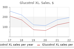
10 mg glucotrol xl order visaNote that the epithelium of the glands in the appendix is much like diabetes mellitus erectile dysfunction 10 mg glucotrol xl generic with amex that of the massive gut treatment of diabetes type 2 glucotrol xl 10 mg cheap on line. Most of the epithelial cells comprise mucin, hence the light appearance of the apical cytoplasm. The lamina propria, as noted, is heavily infiltrated with lymphocytes, and the muscularis mucosae at the base of the glands is tough to recognize (arrows). At the identical degree, the circular layer of the muscularis externa thickens to turn out to be the inner anal sphincter. The external anal sphincter is formed by the striated muscles of the pelvic ground. Mucosa characteristic of the massive gut (colorectal zone) is seen on the higher left of the micrograph. This area is the higher part of the anal canal, and the intestinal glands are the same as those current within the colon. This space known as the anal transitional zone is examined at greater magnification in the bottom left determine. The right rectangular area consists of the stratified squamous epithelium (StS) of the skin within the squamous zone of the anal canal and is examined at larger magnification within the backside right figure. Between the 2 diamonds within the rectangular areas shown is epithelium of the lower a half of the anal canal. Characteristically, the lamina propria incorporates massive numbers of lymphocytes (Lym), notably so in the area marked. A greater magnification of the stratified columnar epithelium (StCol) and stratified cuboidal epithelium (StC) discovered within the transition zone is shown within the inset. The last change in epithelial type that happens on the squamous zone of the anal canal is proven here. Again, quite a few lymphocytes (Lym) are in the underlying connective tissue, and a lot of have migrated into the epithelium within the nonkeratinized area. It is located in the upper right and partially within the higher left quadrants of the abdominal cavity, protected by the rib cage. The liver is anatomically divided by deep grooves into two large lobes (the proper and left lobes) and two smaller lobes (the quadrate and caudate lobes;. This anatomic division has solely topographic significance as a outcome of it relates lobes of the liver to different stomach organs. Division into functional or surgical segments that correspond to the blood provide and bile drainage is more clinically important. In the embryo, the liver develops as an endodermal evagination from the wall of the foregut (specifically the location that will turn into the duodenum) to type the hepatic diverticulum. The diverticulum proliferates, giving rise to the hepatocytes, which turn into arranged in mobile (liver) cords, thus forming the parenchyma of the liver. An outgrowth from the widespread bile duct types the cystic diverticulum that gives rise to the gallbladder and cystic duct. Liver Physiology Many circulating plasma proteins are produced and secreted by the liver. The liver performs an necessary position within the uptake, storage, and distribution of each nutrients and vitamins from the bloodstream. In addition, the liver degrades or conjugates quite a few poisonous substances and medicines, but it could be overwhelmed by such substances and damaged. The liver can also be an exocrine organ; it produces a bile secretion that contains bile salts, phospholipids, and cholesterol. Several vitamins are taken up from the bloodstream and are then stored or biochemically modified by the liver. They embrace: � falciform ligament ligamentum teres terminal hepatic venule (central vein) vitamin A (retinol), an essential vitamin in imaginative and prescient. This diagram exhibits the gross view of the diaphragmatic and visceral surfaces of the liver, with labeled anatomic landmarks discovered on both surfaces. The enlarged cross-sectional area of the liver (bottom) reveals the general microscopic group of the liver into lobules. Note the presence of hepatic portal triads on the periphery of each lobule, with the terminal hepatic venule (central vein) in the heart of the lobule. The circulating plasma proteins produced by the liver embody: � � albumins, that are concerned in regulating plasma quantity and tissue fluid stability by maintaining the plasma colloid osmotic pressure. The liver also produces small quantities of different plasma lipoproteins, such as low-density Vitamin A is the precursor of retinal, which is required for the synthesis of rhodopsin within the eye. The liver performs a serious position in the uptake, storage, and maintenance of circulating ranges of vitamin A. When the vitamin A levels within the blood decrease, the liver mobilizes its storage sites in the hepatic stellate cells (see pages 634�635). Vitamin D is acquired from dietary vitamin D3 and is also produced in the pores and skin during publicity to ultraviolet gentle by conversion of 7-dehydrocholesterol. The liver performs an essential function in vitamin D metabolism by changing vitamin D3 to 25-hydroxycholecalciferol, the predominant type of circulating vitamin D. Further conversion takes place within the kidney to 1,25-hydroxycholecalciferol, which is 10 occasions extra active than vitamin D3. Vitamin D is important for improvement and growth of the skeletal system and enamel. Deficiency of vitamin D is related to rickets and problems of bone mineralization. Vitamin K deficiency is associated with hypoprothrombinemia and bleeding disorders. It synthesizes almost all of the proteins concerned in iron transport and metabolism, together with transferrin, haptoglobin, and hemopexin. The association of the protein with the lipid-containing core makes the complicated sufficiently hydrophilic to remain suspended in the plasma. Lipoproteins serve quite so much of capabilities in cellular membranes and within the transport and metabolism of lipids. The lipoprotein complexes cross to the Golgi, where secretory vesicles containing electron-dense lipoprotein particles bud off and are then launched on the cell surface bordering the perisinusoidal house to attain the bloodstream. Several hormones, such as estrogen and thyroid hormones, regulate the secretion of lipoproteins. These lipoproteins differ in chemical composition and can be isolated from plasma according to their flotation properties, from largest and least dense to smallest and most dense. Chylomicrons, the lightest of all lipoproteins, are made only within the small gut. Their major operate is to transport the big quantity of absorbed fats to the bloodstream. Their function is to transport many of the triglycerides from the liver to different organs. In liver biopsy specimens from these people, large lipid droplets occupy a lot of the hepatocyte cytoplasm.
Glucotrol xl 10 mg buy low priceThe different virilising condition that happens in being pregnant diabetic diet sample menus purchase glucotrol xl 10 mg with visa, theca-lutein cyst diabetic diet oranges trusted glucotrol xl 10 mg, occurs when high ranges of human chorionic gonadotrophin are current because of trophoblastic disease or a quantity of pregnancy; 30 per cent of women could have some degree of virilisation. Blood exams along with androgen testing if the cycles are thought-about to be anovulatory: prolactin ranges; thyroid function. Transvaginal ultrasound scan of ovaries: polycystic ovary pattern; ovarian tumour or cysts. Computed tomography is preferable as it gives better resolution than magnetic resonance imaging, if an adrenal tumour is suspected. Retrograde venous catheterisation may be required to decide the placement (ovary vs. If it has no effect after three months, it should be discontinued, and it could cause rash or worsen acne. Finally, metformin improves peripheral insulin sensitivity and has been proven to have a task, though studies disagree over its advantages in particularly treating hirsutism. Revised 2003 consensus on diagnostic standards and long-term health risks related to polycystic ovary syndrome. Evaluation of ovarian functionality after a dietary remedy in obese ladies with polycystic ovary syndrome. Bleaching, shaving, chemical washes, and mechanical depilation will bodily remove unwanted hairs; laser therapy is pricey but everlasting. The newer progestogens cyproterone acetate (Dianette) and drosperinone (Yasmin) have anti-androgenic results and are best; however, they carry the next risk of venous thromboembolism in comparison with earlier technology drugs. Aldosterone antagonist spironolactone and androgen antagonist flutamide compete with circulating androgens and are second- and third-line choices. Finasteride (a 5a reductase inhibitor lowering peripheral conversion of testosterone to dihydrotestosterone) has also been used. All three reduce libido and trigger feminisation of a male fetus, and therefore careful contraception is essential. Rarely, this type of flushing may be related to scombroid meals poisoning from ingesting spoiled or decaying mackerel or tuna. Drug-associated flushes are associated with: Vasodilators � nitroglycerine, prostaglandins. Fermented alcoholic drinks (beer, sherry) that may contain tyramine or histamine. Harlequin syndrome � hemifacial flushing and sweating with or without warmth and anhidrosis of the contralateral limbs. Definition Hot flushes are episodes of redness of the pores and skin together with a sensation of heat or burning moving down the physique from the face, scalp, and neck, and fewer regularly, the higher trunk and stomach. They may be related to sweats, chills, or vasomotor symptoms similar to palpitations. These assaults are transient, lasting between seconds to minutes, which differentiates them from the persistent erythema of photosensitivity or acute contact dermatitis. Repeated flushing over a time period can result in telangiectasia and sometimes to classical rosacea of the face. A hot flush is related to a rise in core body temperature and pulse price. It is adopted by a decline in temperature and profuse perspiration over the realm of the flush distribution. Mastocytomas � benign proliferative problems of the reticuloendothelial system owing to a hyperplastic somewhat than a neoplastic process. This condition could also be related to headache, shortness of breath, wheezing, palpitations, belly ache, diarrhoea, and syncope. This type of flushing lasts greater than half-hour, unlike the carcinoid or menopausal flush, which lasts less than 10 minutes. Whether the flushing is patchy or confluent in distribution will help to distinguish the signs of menopausal scorching flushing from other dermatological causes similar to dermatitis. Identification of exacerbating or relieving elements may help to develop methods to higher manage and avoid causes of flushing. In particular, avoidance of certain foods can cut back carcinoid flushing and avoidance of alcohol can cut back flushing secondary to mastocytosis and medullary thyroid carcinoma. A useful diagnostic for figuring out causative brokers can be to get the affected person to complete a 2-week food diary to determine whether or not sure meals cause signs. This can then be adopted by a trial period of exclusion of suspected foods to see whether symptoms resolve. However, it is essential to ensure the patient has had a food regimen free of substances which will compound these outcomes previous to testing. Chromogranin-A, substance P, and neurokinin A are other exams helpful in figuring out the non-endocrine causes of flushing, although not requested by a basic gynaecologist. Deep respiratory � paced stomach respiration and muscle contraction relaxation of all muscle groups. Diagnostic or metabolic work-up Pharmacological measures Once the underlying reason for hot flushes has been recognized, using pharmacological measures to control the symptoms can be considered. If symptoms are mild, then way of life modifications ought to be the mainstay of treatment. The immune hydrops is more frequent in growing nations, while the non-immune selection is extra common in developed nations. Mirror syndrome can occur at any time in the course of the antenatal interval and should persist postpartum, which could be life threatening. However, intervention that ends in reversal of fetal hydrops can also reverse the maternal disorder. Spontaneous resolution of mirror syndrome might occur after spontaneous decision of fetal hydrops related to parvovirus B19 and after fetal dying. Fetomaternal haemorrhage happens at placental separation and sensitises the mom. Other procedures during being pregnant that may sensitise the mother are abruptio placentae, external cephalic model, and amniocentesis. In subsequent pregnancies, the fetal purple cells are destroyed by the maternal IgG antibody that can cross the placenta. In mild instances, the fetus has anaemia and haemolytic disease of the new child, but in extreme instances it develops hydrops fetalis. Widespread use of Rh-D immunoglobulin has dramatically decreased the prevalence of Rh-D alloimmunisation and related hydrops. However, rhesus isoimmunisation continues to be the only most common explanation for immune hydrops fetalis in creating countries. This should be carried out within the first instance for the relevant antigen when maternal red cell antibodies are present. Once significant antibodies are detected, levels should be measured each four weeks as much as 28 weeks of gestation and then each 2 weeks till supply. The mechanism for fluid collection in these fetuses could involve obstruction or incomplete formation of the lymphatic system within the neck or stomach, resulting in lymphatic dysplasia.
Buy glucotrol xl 10 mg visaThe sensitivity of transabdominal ultrasound scans is much less correct especially for posterior placenta praevia diabetes symptoms 1 year old glucotrol xl 10 mg purchase fast delivery, whereas a transvaginal ultrasound is more effective in delineating the decrease edge of the placenta diabetes insipidus urine osmolarity glucotrol xl 10 mg buy mastercard. An anterior placenta praevia in a woman with a previous caesarean section should alert the clinician to the potential of a placenta accreta. The best predictive issue for abruption is a historical past of a previous abruption, with a 4 per cent risk for one to a 20�25 per cent threat for two previous abruptions. Urethral vaginal Spontaneous rupture of the uterus in a nulliparous girl with no threat elements has also been described but is fortuitously uncommon. Induction of labour with prostaglandins with subsequent stimulation with Syntocinon will increase the chance. Anal (need to be excluded) Traumatic causes During pregnancy the genital tract will increase in vascularity, and so trauma to any par of the genital tract may cause important bleeding. It ought to be thought-about in all women but particularly in those with the next danger components: laceration following fall; sexual assault; international body; traumatic use of intercourse toys, from circumferential harm at the introitus to a tear on the vaginal fornices as a end result of deep penetrative injury; the latter may cause profound bleeding. When these vessels are under the presenting half in the region of the interior os the time period vasa praevia is used. The earliest indicators of hypovolaemia are an increase in respiratory price, a delay in capillary refill time, and an abnormality of the cardiotocograph. Hypotension could also be a late sign up young wholesome girls who compensate well initially. Women with these signs should be stabilised and immediately transferred to a consultant-led unit or any facility the place an emergency delivery and transfusion of the mother could be performed. All units should have a massive obstetric haemorrhage protocol which incorporates contacting the haematologist on name for advice relating to the use of blood merchandise. Sudden onset of painless, bright pink vaginal bleeding may suggest placenta praevia. Women with placenta praevia may have a few episodes of warning bleeds before a major bleed, and admission for remark must be considered. Sudden onset of a extreme steady stomach pain related to darker bleeding could recommend placental abruption. If the bleeding is associated with a thick mucoid plug, this can be a heavy show associated with the onset of labour. These vessels could rupture with both spontaneous or synthetic rupture of membranes, causing vital fetal haemorrhage and even dying. A soft, non-tender stomach with a high unengaged presenting part or irregular lie is suggestive of placenta praevia. The uterus may be markedly tender throughout or localised to where the placental abruption has occurred. It may be due to a quantity of delicate abruptions (separations) on the periphery of the placenta. It is important that the huge obstetric haemorrhage protocol is activated, appropriate blood merchandise are used with haemotological advice, and energetic management of the third stage of labour and early detection and remedy of a consumptive coagulopathy are accomplished to stop this from occurring. If the woman has not been booked in the index pregnancy, an ultrasound scan should precede a digital or a speculum vaginal examination to exclude a serious placenta praevia, as examination might irritate the bleeding. A speculum examination may assist diagnose local causes of vaginal bleeding in addition to assist assess cervical dilatation. As mentioned earlier a transvaginal ultrasound is more accurate than a transabdominal ultrasound. Ultrasound may not be helpful in an early abruption where the analysis is usually a clinical one. This is by definition a retrospective analysis of amenorrhoea of 1-year length as a outcome of failure of ovarian operate. Any vaginal bleeding that happens after 6 months of amenorrhoea from presumed menopause ought to be handled as suspicious. Investigations ought to be directed to decide the trigger of bleeding relying on the age of the girl. If she is overweight, the risk is increased to 18 per cent, and to 21 per cent if diabetic. The massive obstetric protocol should be activated, with urgent anaesthetic and obstetric input on the marketing consultant level. Investigations to enable additional management Ideally, a Kleihauer test should be performed in rhesus-negative girls to quantify fetomaternal haemorrhage in order that the suitable dose of anti-D may be calculated. Placental abruption could be the scientific presentation of a girl with extreme pre-eclampsia. It is necessary to consider this in all women with abruption and the Common causes these are summarised in Box 1 and regarded in additional detail beneath. Consequently, most clinicians would use these preparations with warning and after counselling concerning the theoretical increased threat of oestrogen levels that can contribute to breast most cancers recurrence. Atrophic endometritis Endometrial inflammation and thinning that happens as a end result of oestrogen deficiency is named atrophic endometritis. It might result in postmenopausal recognizing and even bleeding, particularly in hypertensive girls. This is a analysis of exclusion, arrived at after the more sinister pathological causes of postmenopausal bleeding within the uterus have been excluded. Endometrial polyps are often inflammatory, but could often have hyperplastic or neoplastic adjustments of the masking endometrium. Uterine polyps can also be of fibroid origin and are much more widespread in a fibroid uterus, although they rarely turn into sarcomatous. Intrauterine polyps could additionally be recognized on transvaginal ultrasound or seen simply as thickened endometrium. Atrophic vaginitis Atrophic vaginitis is attributable to non-specific vaginal irritation and excessive thinning of vaginal epithelium on account of oestrogen deficiency. Because of atrophic adjustments, even the slightest of trauma from intercourse or dabbing oneself dry may lead to bleeding. Apart from postmenopausal bleeding, it may also be associated with dyspareunia, vaginal pruritus, dryness, and pain. It is a standard condition, which impacts up to forty five per cent of postmenopausal ladies. This condition is definitely treated and prevented by the native application of oestrogen lotions. As topical oestrogens have restricted systemic absorption, they can be utilized in an unopposed nature in women with a uterus for a restricted time span. Factors aiding the choice to undertake a hysterectomy will be presence of signs, the age, and the final medical situation of the postmenopausal woman. It should be borne in thoughts that, in postmenopausal women, levels of circulating oestrogens are low. The improvement of hyperplasia may be reflective of steady oestrogen stimulation with either exogenous or endogenous oestrogens.
Glucotrol xl 10 mg overnight deliveryNote that blood sugar 380 cheap glucotrol xl 10 mg mastercard, in distinction to the distal straight tubules diabetes insipidus head trauma glucotrol xl 10 mg with visa, the proximal straight tubules show a brush border and have a larger exterior diameter, with many displaying a star-shaped lumen. The renal corpuscle seems as a spherical construction whose periphery consists of a thin capsule that encloses a slim clear-appearing house, the urinary space (asterisks), and a capillary tuft or glomerulus that appears as a large cellular mass. The visceral layer consists of cells referred to as podocytes (Pod) that lie on the outer floor of the glomerular capillary. Except where they clearly line the urinary space, because the labeled cells do in determine on left, podocytes could also be difficult to distinguish from the capillary endothelial cells. To complicate matters, the mesangial cells are additionally a element of the glomerulus. In basic, nuclei of podocytes are larger and stain much less intensely than do the endothelial and mesangial cell nuclei. In determine on right, both the vascular pole and the urinary pole of the renal corpuscle are evident. The vascular pole is characterised by the presence of arterioles (A), certainly one of which is entering or leaving (double-headed arrow) the corpuscle. The afferent arteriole possesses modified clean muscle cells with granules, the juxtaglomerular cells (not evident in this figure). Here, the urinary house of the renal corpuscle continues into the lumen of the proximal tubule, and the lining cells change from simple squamous to easy cuboidal or low columnar with a brush border. The medulla contains the thick straight segments of proximal and distal tubules, together with their thin segments, the accumulating ducts, and the blood vessels that run in parallel with them. These constructions function because the countercurrent multiplier and countercurrent trade systems that, ultimately, produce hypertonic urine. The ultimate urine drains from the papillary ducts (of Bellini) into calyces that then empty into the renal pelvis. This area incorporates proximal and distal thick segments, thin segments, and medullary collecting ducts. All of the tubules are parallel, and all are cut in cross-section; thus, they current round profiles. The proximal straight tubules (P) show typical star-shaped lumina and a brush border (or the fragmented apical cell floor from which the comb border has been partially broken). These tubules have outside diameters which are usually bigger than these of the distal straight tubules (D). As mentioned beforehand and as shown here, the distal straight tubules display a larger variety of nuclei than do comparable segments of proximal straight tubule cells. Note, additionally, that the lumen of the distal tubule is more rounded and the apical floor of the cells is sharper. The cells forming the accumulating ducts are cuboidal and smaller than these of proximal tubules; thus, additionally they show a comparatively larger number of nuclei than do comparable segments of proximal tubule cells. Finally, boundaries between the cells that constitute the amassing ducts are usually evident (asterisks); this serves as one of the dependable options for the identification of collecting ducts. The thin segments (T) have the thinnest partitions of all renal tubules seen within the medulla. They are fashioned by a low cuboidal or simple squamous epithelium, as seen right here, and the lumina are comparatively large. Occasionally, a bit includes the region of transition from a thick to a skinny section and may be acknowledged even in a cross-section through the tubule. One such junction is evident on this figure (the tubule with two arrows within the lumen). On one side, the tubule cell (left-pointing arrow) is attribute of the proximal segment; it possesses a particular brush border. The different side of the tubule (right-pointing arrow) is composed of low cuboidal cells that resemble the cells forming the thin segments. In addition to the uriniferous tubules and amassing ducts, there are many other small tubular constructions in this figure. The pyramid is a conical structure composed principally of medullary straight tubules, ducts, and the straight blood vessels (vasa recta). The dashed line at the left of the micrograph is placed on the junction between cortex and medulla; thus, it marks the bottom of the pyramid. The pyramid is considerably distorted in this specimen, as evidenced by regions of longitudinally sectioned tubules, decrease left, and cross-sectioned and obliquely sectioned tubules in other areas. In impact, part of the pyramid was bent, thus the change within the plane of section of the tubules. The apical portion of the pyramid (arrowhead), often recognized as the renal papilla, is lodged in a cup- or funnel-like construction referred to as the calyx. It collects the urine that leaves the tip of the papilla from the papillary ducts (of Bellini). Although not evident on the low magnification shown here, the boundary between the columnar epithelium overlaying the papilla and the transitional epithelium overlaying the inside surface of the calyx is marked by the diamonds. They are lined with transitional epithelium (urothelium), an impervious layer that traces the urinary excretory passages from the renal calyces via the urethra. The ability of this epithelium to turn out to be thinner and flatter permits all of those passages to accommodate to distension by the urine. The epithelium rests on a dense collagenous lamina propria, which in flip, rests on an internal longitudinal and an outer round layer of easy muscle. Regular peristaltic contractions of this muscle contribute to the flow of urine from the kidney to the urinary bladder. Note that the ureters are situated behind the peritoneum of the stomach cavity in their course to the bladder. Thus, a serosa (Ser) may be found overlaying a portion of the circumference of the tube. Also, due to contraction of the sleek muscle of the muscularis, the luminal surface is characteristically folded, thus creating a star-shaped lumen. The transitional epithelium and its supporting connective tissue represent the mucosa (Muc). However, the outer longitudinal layer is current solely on the lower end of the ureter. In a cross-section by way of the ureter, the inside and outer smooth muscle layers are minimize in cross-section, whereas the center circular layer of the muscle cells is minimize longitudinally. The basal cells are the smallest, and typically, the nuclei appear crowded because of the minimal cytoplasm of every cell. The intermediate cells seem to encompass a quantity of layers and are composed of cells bigger in dimension than the basal cells but smaller than the floor cells. The wall of the ureter from the oblong space in the orientation micrograph is examined at greater magnification in this determine. One can immediately acknowledge the thick epithelial lining, which appears distinct and sharply delineated from the remainder of the wall. Note that the nuclei appear as round profiles, indicating that the muscle cells have been cross-sectioned. As in most distensible hole viscera that vacant their contents through a slender aperture, the smooth muscle within the wall of the urinary bladder is less regularly arranged than the description signifies, permitting contraction to cut back the amount comparatively evenly throughout the bladder.
Diseases - Adrenogenital syndrome
- Leukocytoclastic angiitis
- Mental retardation hypotonia skin hyperpigmentation
- Yersiniosis
- Zunich Kaye syndrome
- Acromesomelic dysplasia Brahimi Bacha type
- Infantile onset spinocerebellar ataxia
10 mg glucotrol xl generic mastercardThis photomicrograph is from the lung of an individual in the early phases of acute pneumonia (inflammation of the lung) metabolic disease ketonuria glucotrol xl 10 mg discount line. Note that the air areas are crammed with exudate containing white blood cells (mainly neutrophils) diabetes insipidus electrolyte levels cheap glucotrol xl 10 mg mastercard, red blood cells, and fibrin. The capillaries in the alveolar septum are enlarged and congested with purple blood cells. The lower proper nook shows early group of the intra-alveolar exudate; observe that the growing fibrin network contains entrapped neutrophils and a quantity of other pink blood cells. Three principal features of the respiratory system are air conduction, air filtration, and gas trade (respiration). The higher a half of the respiratory system (nasal cavities, paranasal sinuses, nasopharynx, and oropharynx) develops from the primitive oral cavity. The lower a part of the respiratory system (larynx, trachea, bronchi with their divi- sions, and lungs) develops from the ventral evagination of the foregut endoderm. The conducting portion of the respiratory system consists of the upper a part of the respiratory system, larynx, trachea, bronchi, and most of the bronchioles (up to the terminal bronchioles). The respiratory portion accommodates the respiratory bronchioles, alveolar ducts, alveolar sacs, and alveoli. The respiratory area is lined by the respiratory mucosa that incorporates ciliated, pseudostratified columnar epithelium. It possesses a wealthy vascular community in the lamina propria, in addition to quite a few mucus- and serous-secreting glands. The olfactory area located at the roof of the nasal cavity is lined by a pseudostratified olfactory epithelium with out goblet cells. Olfactory epithelium is composed of olfactory receptor cells (bipolar neurons), supporting cells, brush cells, and basal cells. Olfactory receptor cells possess apical immotile cilia, which comprise G protein�coupled receptors involved within the olfactory transduction pathway. It contains vocal folds that control the circulate of air through the larynx and vibrate to produce sound. The larynx is lined by the respiratory mucosa, apart from the luminal floor of the vocal cords, which is covered by a stratified squamous epithelium. The wall of the trachea consists of 4 layers: mucosa (composed of a ciliated, pseudostratified epithelium resting on a thick basement membrane), submucosa (dense irregular connective tissue), cartilaginous layer (composed of Cshaped hyaline cartilages), and adventitia (binds the trachea to adjoining structures). Bronchi are lined by respiratory mucosa with the identical cellular composition as the trachea. The smallest conducting terminal bronchioles are lined with a easy cuboidal epithelium containing Clara cells. These Respiratory System cells produce a surface-acting agent that stops airway collapse. Respiratory bronchioles are the primary part of the bronchial tree that allows fuel change. Pulmonary circulation delivers blood through branches Their septa are the places for gasoline exchange between the air and the blood. Type I alveolar cells are extremely skinny squamous cells that line 95% of the alveolar floor and form the barrier between the air area and the septal wall. It consists of a thin layer of surfactant, a kind I epithelial cell with its basal lamina, and a capillary endothelial cell with its basal lamina. Alveolar and septal macrophages are current in alveolar air areas and septal connective tissue, respectively. Blood is collected by pulmonary venous capillaries that finally form the pulmonary veins. Bronchial circulation, through bronchial arteries, supplies the partitions of the bronchi, bronchioles, and the remaining connective tissue of the lung. Autonomic nerves comply with the branches of pulmonary arteries and innervate the graceful muscle of blood vessels, the bronchial tree, and the respiratory mucosa. Its pseudostratified epithelium is thicker than that of nonsensory epithelium, and it serves as the receptor for odor. Olfactory epithelium consists of olfactory cells, supporting (sustentacular) cells, basal cells, and brush cells. The apex of the cell is expanded into the olfactory vesicle from which nonmotile cilia, the actual receptors, lengthen into surface secretions. The base of the cell tapers into an axonal course of that enters the lamina propria and joins axons from other receptor cells to type the olfactory nerve. Large, cuboidal Schwann cells are a distinguished feature of those axons, giving the nerve an uncommon appearance. They attach to the receptor cells through adhering junctions and provide mechanical and metabolic support to the olfactory cells. Basal cells are stem cells from which olfactory and supporting cells differentiate. Brush cells are the identical cell sort that occurs in nonsensory respiratory epithelium. These are tubuloalveolar serous glands whose watery secretion serves as a entice and solvent for odorant substances and continuously washes the olfactory floor. This low-magnification orientation micrograph shows part of the wall of the nasal cavity. The olfactory mucosa is immediately attached to the bone tissue; no submucosa is current. In this specimen, nonetheless, the mucosa is separated from the bone tissue due to shrinkage, a regularly encountered artifact. Note that the adjoining respiratory mucosa lacks the nerves and reveals a relative paucity of glands. The supporting cell has a cylindrical form and extends from the basement membrane by way of the complete thickness of the epithelium. Careful examination of the nuclei of those bipolar neuronal cells reveals that they comprise extra euchromatin than the nuclei of the supporting cells and sometimes exhibit several nucleoli. Note that the olfactory mucosa in distinction to respiratory mucosa lacks goblet cells. The duct components lengthen from the secretory portion of the gland beginning in close proximity to the overlying epithelium (arrowhead) and pass immediately by way of the epithelium to deliver their secretions at the surface. The nuclei current within the olfactory nerves represent Schwann cell nuclei (ScC). It consists of a cartilaginous framework to which both extrinsic and intrinsic muscle tissue are hooked up and a mucosal floor that varies in character from pseudostratified to stratified squamous in areas topic to abrasion by the air stream. The muscle tissue transfer certain cartilages with respect to others, thus rising or lowering the opening of the rima glottis and increasing or reducing the strain on the vocal folds (cords). In this fashion, vibrations of different wavelengths are generated in the passing air, and sound is produced. The vocal folds are ridge-like buildings which are oriented in an anteroposterior (ventral-dorsal) direction.
Generic 10 mg glucotrol xl otcThe rectangular space within the figure under is taken into account at larger magnification right here diabetes type 1 hypersensitivity glucotrol xl 10 mg purchase on line. The surface cells include an apical cup of mucous material that usually appears empty in an H&E�stained paraffin section diabetes prevention handout cheap glucotrol xl 10 mg line. The intestinal absorptive cells also possess a striated border, which is shown in Plate 60. It is the longest element of the alimentary canal, measuring over 6 m, and is split into three segments: duodenum (25 cm), jejunum (2. The first portion, the duodenum, receives a partially digested bolus of meals (chyme) from the abdomen, in addition to secretions from the abdomen, pancreas, liver, and gallbladder that include digestive enzymes, enzyme precursors, and other products that assist digestion and absorption. The small gut is characterised by plicae circulares, everlasting transverse folds that contain a core of submucosa, and villi, finger-like and leaf-like projections of the mucosa that stretch into the lumen. Microvilli, multiple finger-like extensions of the apical surface of every intestinal epithelial cell (enterocyte), additional enhance the floor for absorption of metabolites. They comprise the stem cells and creating cells that may in the end migrate to the floor of the villi. Enterocytes not solely take in metabolites digested within the intestinal lumen but additionally synthesize enzymes inserted into the membrane of the microvilli for terminal digestion of disaccharides and dipeptides. Both longitudinal (L) and round (C) layers of the muscularis externa can be distinguished. Although plicae circulares are discovered in the wall of the small gut, including the duodenum, none is included on this photomicrograph. A distinctive function of the intestinal mucosa is the presence of finger-like and leaf-like projections into the intestinal lumen, called villi. Most of the villi (V) proven here display profiles that correspond to their description as finger-like. The dashed line marks the boundary between the villi and the intestinal glands (also referred to as crypts of Lieberk�hn). These are branched tubular or branched tubuloalveolar glands whose secretory elements, proven at greater magnification within the figure beneath, include columnar epithelium. The lamina propria additionally contains elements of free connective tissue and isolated easy muscle cells. The bases of the intestinal crypts comprise the stem cells from which all the different cells of the intestinal epithelium arise. The granules comprise lysozyme, a bacteriolytic enzyme thought to play a task in regulating intestinal microbial flora. The major cell type within the intestinal crypt is a comparatively undifferentiated columnar cell. These cells are shorter than the enterocytes of the villus floor; they often bear two mitoses earlier than they differentiate into absorptive cells or goblet cells. Also present in the intestinal crypts are some mature goblet cells and enteroendocrine cells. The histologic options of the duodenal mucosa are proven at higher magnification right here. They have a striated border that might be seen at larger magnification in Plate 60; their elongate nuclei are positioned within the basal half of the cell. Goblet cells are readily identified by the presence of the apical mucous cup, which appears empty. Most of the dark spherical nuclei also seen within the epithelial layer masking the villi belong to lymphocytes. The villi are more finger-like than leaf-like and are covered largely with absorptive columnar epithelial cells (enterocytes), although goblet cells and enteroendocrine cells are also present. The stem cells for all of those cells and the Paneth cells that secrete the antibacterial enzyme lysozyme are discovered deep in the intestinal gland. These folds or ridges are largely arranged with their long axis at roughly right angles to the longitudinal axis of the gut; due to this fact, the plicae circulares shown listed under are reduce in cross-section. The shortening is taken into account to be because of the contraction of easy muscle cells in the villi. Lacteals are lymphatic capillaries that start within the villi and carry sure absorbed dietary lipids and proteins from the villi to the larger lymphatic vessels of the submucosa. The glands are surrounded by cells of the lamina propria; the villi are surrounded by space of the intestinal lumen. The lamina propria with its lacteal occupies a central position within the villus; the lumen occupies the central position of the gland. Studies of enzymatically isolated preparations of mucosa show that the bases of the glands are often divided into two or three finger-like extensions resting on the muscularis mucosae. These processes partially delimit the basal-lateral intercellular areas (asterisks) that are dilated, as may be seen right here, during energetic transport of absorbed substrates. In this specimen, the nucleus of almost each goblet cell is simply at the base of the cup, and a skinny cytoplasmic strand (not always evident) extends to the level of the basement membrane. Part of the plica circularis marked by the bracket in the determine above is shown at higher magnification. Some of the glands are reduce longitudinally; some are minimize in cross-section; many of the villi have been cut longitudinally. The darkish band at the base of the striated border is as a result of of the terminal internet of the cell, a layer of actin filaments that extends throughout the apex of the cell to which the actin filaments of the cores of the microvilli attach. The nuclei of the enterocytes have primarily the identical shape, orientation, and marking characteristics. The enterocytes relaxation on a basal lamina not evident in H&E� stained paraffin sections. Some, nevertheless, are emphasized; specifically, villi in the ileum are extra incessantly leaf-like, and lymphatic tissue within the lamina propria is organized into small and enormous nodes that are present in nice number on the antimesenteric side of the ileum. The stem cells are restricted to the bottoms of the mucosal glands, and the zone of cell replication is restricted to the lower half of the gland. Just inner to the submucosa is the mucosa; exterior to the muscularis externa is the serosa. The mucosa reveals several longitudinally sectioned villi (V), which have been labeled, and different unlabeled villi, which may be recognized easily on the premise of their appearance as islands of tissue utterly surrounded by the space of the lumen. They are, in fact, not islands as a outcome of this appearance is due to the plane of section that slices fully by way of a few of the villi obliquely or in cross-section, thereby isolating them from their base. There are about eight to 10 projections of tissue into the intestinal lumen which are considerably larger than the villi. As famous above, plicae usually have round orientation, but they could journey in a longitudinal direction for short distances and will department. In addition, even if all of the plicae are arranged in a circular method, if the part is considerably oblique, the plicae shall be minimize at an angle, as appears to be the case with a quantity of plicae in this figure. One of the distinctive options of the small gut is the presence of single and aggregated lymph nodules within the intestinal wall. Isolated nodules of lymphatic tissue are widespread within the proximal end of the intestinal canal.
Glucotrol xl 10 mg safeThe dashed line marks the boundary between the villi and the everyday intestinal glands (crypts of Lieberk�hn) type 2 diabetes definition cdc discount 10 mg glucotrol xl mastercard. These are branched tubular glands whose secretory element consists of columnar cells blood glucose numbers chart cheap glucotrol xl 10 mg line. This intestinal stem cell area of interest (zone of cell replication) is restricted to the lower one-half of the gland and contains highly proliferative intermediate cells (as beforehand explained) and cells at various levels of differentiation. A cell destined to turn out to be a goblet cell or absorptive cell normally undergoes a number of extra divisions after it leaves the pool of stem cells. The epithelial cells migrate upward in the intestinal gland onto the villus the place they endure apoptosis and slough off into the lumen. Autoradiographic studies have proven that the renewal time for absorptive and goblet cells within the human small gut is four to 6 days. Enteroendocrine cells and Paneth cells are also derived from the stem cells on the base of the intestinal gland. They live for about four weeks and are then changed by differentiation of a close-by "dedicated" cell in the intestinal gland. As mentioned within the chapter on epithelial tissue (page 146), expression of the transcription issue Math1 appears to determine the destiny of differentiating cells in the intestinal stem cell niche. The colon is additional subdivided on the premise of its anatomic location into ascending colon, transverse colon, descending colon, and sigmoid colon. Local contractions displace intestinal contents both proximally and distally; this type of contraction is known as segmentation. They serve to circulate the chyme locally, mixing it with digestive juices and shifting it into contact with the mucosa for absorption. Peristalsis, the second kind of contraction, includes coordinated motion of each circular and longitudinal muscle layers and strikes the intestinal contents distally. Haustra coli that are visible sacculations between the teniae coli on the external floor of the cecum and colon. Omental appendices that are small fatty projections of the serosa, observed on the outer surface of the colon. Serosa the serosa of the elements of the small gut that are situated intraperitoneally within the belly cavity corresponds to the final description firstly of the chapter. Mucosa the mucosa of the large intestine has a "easy" floor; neither plicae circulares nor villi are current. The glands consist of simple columnar epithelium, as does the intestinal surface from which they invaginate. Examination of the luminal floor of the big gut on the microscopic level reveals the openings of the glands, that are arranged in an orderly pattern. The morphology of absorptive cells is basically similar to that of the enterocytes of the small gut. Elimination of semisolid to strong waste materials is facilitated by the big quantities of mucus secreted by the quite a few goblet cells of the intestinal glands. Goblet cells are more numerous within the large gut than within the small intestine. The mucosal epithelium of the massive gut contains the identical cell types as the small intestine except Paneth cells, that are usually absent in humans. This photograph exhibits the outer (serosal) surface (left) and inner (mucosal) floor (right) of the transverse colon. The easy mucosal floor exhibits semilunar folds (arrows) shaped in response to contractions of the muscularis externa. The ratio decreases, nevertheless, approaching 1:1, near the rectum, where the number of goblet cells increases. This photomicrograph of an H&E preparation shows the mucosa and a half of the submucosa. The floor epithelium is continuous with the straight, unbranched, tubular intestinal glands (crypts of Lieberk�hn). As the absorptive cells are adopted into the glands, they become fewer in quantity, whereas the goblet cells increase in quantity. The highly mobile lamina propria accommodates quite a few lymphocytes and other cells of the immune system. Comparative electron-microscopic options of regular, hyperplastic, and adenomatous human colonic epithelium. Goblet cells could mature deep in the intestinal gland, even in the replicative zone. They secrete mucus continuously, even to the point where they attain the luminal floor. Here, on the floor, the secretion rate exceeds the synthesis rate, and "exhausted" goblet cells appear within the epithelium. These cells are tall and skinny and have a small number of mucinogen granules in the central apical cytoplasm. An occasionally noticed cell sort, the caveolated "tuft" cell, has additionally been described within the colonic epithelium; nonetheless, this cell could additionally be a form of exhausted goblet cell. The turnover instances of the epithelial cells of the massive intestine are much like those of the small intestine. Senile epithelial cells that reach the mucosal floor undergo apoptosis and are shed into the lumen at the midpoint between two adjoining intestinal glands. Lamina Propria Although the lamina propria of the massive gut contains the same primary parts as the rest of the digestive tract, it demonstrates some additional structural options and greater development of some others. These embody the next: � Collagen table, which represents a thick layer of col- Epithelial Cell Renewal within the Large Intestine All intestinal epithelial cells within the massive gut derive from a single stem cell inhabitants. As within the small intestine, all of the mucosal epithelial cells of the large intestine come up from stem cells situated at the backside of the intestinal gland. The intermediate cell varieties found in the � lagen and proteoglycans that lies between the basal lamina of the epithelium and that of the fenestrated absorptive venous capillaries. This layer is as a lot as 5 m thick within the regular human colon and could be as much as thrice that thickness in human hyperplastic colonic polyps. The collagen desk participates in regulation of water and electrolyte transport from the intercellular compartment of the epithelium to the vascular compartment. Pericryptal fibroblast sheath, which constitutes a well-developed fibroblast inhabitants of regularly replicating cells. Some evidence suggests that the macrophages of the core of the lamina propria in the massive intestine could arise as a terminal differentiation of the pericryptal fibroblasts. The intensive development of the immune system in the colon most likely displays the large number and variety of microorganisms and noxious finish products of metabolism usually current in the lumen. Only lately, utilizing new very selective markers for lymphatic epithelium, researchers have found occasional small lymphatic vessels on the bases of the intestinal glands.
Buy glucotrol xl 10 mg low costMucus diabetes mellitus versi indonesia 10 mg glucotrol xl effective, an acid-protective coating for the abdomen secreted by a number of kinds of mucus-producing cells diabetic diet sample menu 10 mg glucotrol xl buy fast delivery. The mucus and bicarbonates trapped within the mucous layer maintain a impartial pH and contribute to the so-called physiologic gastric mucosa barrier. In addition, mucus serves as a bodily barrier between the cells of the gastric mucosa and the ingested materials within the lumen of the stomach. Intrinsic issue, a glycoprotein secreted by parietal cells that binds to vitamin B12. Lack of intrinsic factor results in pernicious anemia and vitamin B12 deficiency (see Folder 17. In addition, undifferentiated cells that give rise to these cells are additionally current. These are the various cells that constitute the gland: � � � � � Mucous neck cells Chief cells Parietal cells, additionally called oxyntic cells Enteroendocrine cells Undifferentiated grownup stem cells Mucous neck cells are localized in the neck region of the gland and are interspersed with parietal cells. As the name implies, the mucous neck cells are situated within the neck area of the fundic gland. The mucous neck cell is way shorter than the surface mucous cell and contains considerably less mucinogen within the apical cytoplasm. Also, the nucleus tends to be spherical compared with the more prominent, elongated nucleus of the floor mucous cell. The mucous neck cells secrete much less alkaline soluble mucus in contrast with the high-alkaline insoluble or cloudy mucus produced by the floor mucous cell. These mucous neck cells differentiate from stem cells, which reside in the neck region of the fundic gland. Gastric ulcers are current in 95% of sufferers with this syndrome and are six occasions extra prevalent than the duodenal ulcers. Patients with Zollinger-Ellison syndrome may expertise intermittent stomach ache, diarrhea, and steatorrhea (excretion of stool containing a large amount of fat). In addition, surgical excision of the tumor, when possible, removes the source of gastrin production and alleviates symptoms. The basophilia, particularly, allows easy identification of these cells in H&E sections. On contact with the acid gastric juice, pepsinogen is converted to pepsin, a proteolytic enzyme. They are giant cells, generally binucleate, and appear somewhat triangular in sections, with the apex directed toward the lumen of the gland and the bottom resting on the basal lamina. The nucleus is spherical, and the cytoplasm stains with eosin and other acidic dyes. Their size and distinctive staining characteristics enable them to be simply distinguished from different cells within the fundic glands. The cytoplasm of the parietal cell stains with eosin largely due to the intensive quantity of membrane comprising the intracellular canaliculus, tubulovesicular system, mitochondria, and the relatively small number of ribosomes. There is evidence that chromogranin A regulates biosynthesis of dense-core secretory granules, whereas chromogranin B controls sorting and packaging of produced peptides into secretory vesicles. These tumors symbolize uncommon neoplasms of the gastrointestinal tract and pancreas that often secrete hormonally lively brokers, causing distinct scientific syndromes. The appendix is the most common gastrointestinal website of origin for neuroendocrine tumors. The classical instance is the carcinoid syndrome brought on by the discharge of a wide selection of hormonally energetic substances by tumor cells. Symptoms include diarrhea (case by serotonin), episodic flushing, bronchoconstriction, and right-sided cardiac valve disease. The enteroendocrine cells differentiate from the progeny of the same stem cells as all the other epithelial cells of the digestive tract. Enteroendocrine cells are specialised cells present in the mucosa of the digestive tract. They account for lower than 1% of all epithelial cells within the gastrointestinal tract, however as a complete, they collectively constitute the biggest endocrine "organ" within the body. Enteroendocrine cells are also found within the ducts of the pancreas, the liver, and the respiratory system, another endodermal spinoff that originates by invagination of the epithelium of the embryonic foregut. Rather, enteroendocrine cells are distributed singly throughout the gastrointestinal epithelium. Here, enteroendocrine cells, derived from pancreatic buds that additionally come up from the embryonic foregut, type specialized accumulations called endocrine islets of Langerhans (see web page 647). A paracrine hormone differs from an endocrine hormone in that it diffuses domestically to its goal cell as an alternative of being carried by the bloodstream to a target cell. A well-known substance that appears to act as a paracrine hormone within the gastrointestinal tract and pancreas is somatostatin, which inhibits different gastrointestinal and pancreatic islet endocrine cells. Functional Considerations: the Gastrointestinal 579 Other locally lively agents isolated from the gastrointestinal mucosa are neurotransmitters. These agents are released from nerve endings close to the target cell, often the smooth muscle of the muscularis mucosae, the muscularis externa, or the tunica media of a blood vessel. Thus, a particular peptide could additionally be produced by endocrine and paracrine cells and in addition be localized in nerve fibers. In an actively secreting cell, the variety of microvilli within the canaliculi increases, and the tubulovesicular system is decreased considerably or disappears. The membranes of the tubulovesicular system function a reservoir of plasma membrane containing energetic proton pumps. This membrane materials may be inserted into the plasma membrane of the canaliculi to improve their floor space and the number of proton pumps obtainable for acid manufacturing. Numerous mitochondria with advanced cristae and many matrix granules supply the high ranges of power needed for acid secretion. Activation of the gastrin receptor by gastrin, a gastrointestinal peptide hormone, is the major path for parietal cell stimulation (Folder 17. Simultaneously, K inside the canaliculus is transported into the cell in trade for the H ions. Cl ions are additionally transported from the cytoplasm of the parietal cell into the lumen of the canaliculus by Cl channels in the membrane. Simultaneously, K from the canaliculus is transported into the cell cytoplasm in change for the H ions. Transport of K and Cl ions from the parietal cell cytoplasm into the lumen of the canaliculus via activation of K and Cl channels (uniporters) in the plasma membrane. Intrinsic issue is a forty four kDa glycoprotein that complexes with vitamin B12 within the stomach and duodenum, a step essential for subsequent absorption of the vitamin within the ileum. Autoantibodies directed towards intrinsic issue or parietal cells themselves lead to an intrinsic issue deficiency, leading to malabsorption of vitamin B12 and pernicious anemia (see Folder 17. Enteroendocrine cells secrete their products into both the lamina propria or underlying blood vessels. In humans, intrinsic issue is secreted by the parietal cells (chief cells accomplish that in another species).
|
