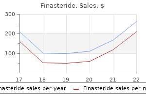
Generic 1 mg finasteride fast deliveryAuerbach A et al: Focal myositis: a clinicopathologic research of 115 cases of an intramuscular mass-like reactive course of hair loss golden retriever buy finasteride 1 mg low price. Fine Capillary Network Eosinophilic Polygonal Cells (Left) In most circumstances of grownup rhabdomyoma hair loss cure kids order finasteride 1 mg otc, the predominant tumor cell is large and polygonal and incorporates outstanding eosinophilic cytoplasm. Tumor cells can also contain cytoplasmic vacuolizations and appear completely clear or demonstrate a retracted "spider cell" morphology. It is characterized by loose bundles and fascicles of spindled eosinophilic cells inside a distinguished myxoid stroma. It often arises in grownup ladies (particularly in the vagina) but can be seen in males and in youngsters. In women, lesions are typically polypoid, as depicted, with overlying squamous epithelium. Prominent Clear Cell Morphology Vacuolated Tumor Cells (Left) Although many of the tumor cells in cardiac rhabdomyoma are closely vacuolated clear cells, some cells are much less vacuolated and present eosinophilic cytoplasm. Of note, perivascular regions are often more mobile than the adjacent looser myxoid zones. Only nuclear expression ought to be counted, as cytoplasmic expression is nonspecific. Tumors with in depth fascicular growth are greatest thought of spindle cell rhabdomyosarcomas. This morphology might simulate strong development in alveolar rhabdomyosarcoma if myxoid stroma is limited. In addition to bigger epithelioid cells, some cells are more elongated and myofiber-shaped and may be referred to as "strap cells" or "tadpole cells. Pseudoalveolar Pattern Pseudoalveolar Pattern (Left) Despite the lack of mobile cohesion in the center of the nests, the peripheral cells typically remain connected to the fibrous septa, an look somewhat resembling alveoli. These cells classically present a peripheral or wreath-like association of nuclei and plentiful eosinophilic cytoplasm. This morphology might result in confusion with Ewing sarcoma, and immunohistochemistry is often useful. To qualify for this designation, this strong progress ought to reflect the majority of the tumor. In some instances, infiltration of skeletal muscle is distinguished and resembles the myoinvasive pattern of lymphoma. Importantly, only nuclear expression counts for these markers, as cytoplasmic staining is nonspecific and should be ignored. Stock N et al: Adult-type rhabdomyosarcoma: evaluation of fifty seven circumstances with clinicopathologic description, identification of 3 morphologic patterns and prognosis. Mentzel T et al: Spindle cell rhabdomyosarcoma in adults: clinicopathological and immunohistochemical evaluation of seven new circumstances. Weaker staining is seen throughout the fascicles of normal skeletal muscle in this image. A fascicular growth pattern continues to be current, however no obvious rhabdomyoblasts are seen in this field. Some smaller nests could present central dyscohesion, imparting a microalveolar look. Note, nonetheless, that the attribute hyalinized stroma can still be appreciated, even at low magnification. It is characterized by clusters and nests of loosely cohesive tumor cells with central areas, harking again to small pulmonary alveoli. In conjunction with the sclerotic stroma, this look might simply result in confusion with sclerosing epithelioid fibrosarcoma. Pleomorphic rhabdomyoblasts are massive polygonal cells with markedly atypical nuclei and plentiful deeply eosinophilic cytoplasm. Pleomorphic Rhabdomyoblasts Pleomorphic Rhabdomyoblasts (Left) Pleomorphic rhabdomyoblasts exhibit a diverse array of styles and sizes. Li G et al: Cytogenetic and real-time quantitative reverse-transcriptase polymerase chain reaction analyses in pleomorphic rhabdomyosarcoma. The diploma of atypia can be extreme in some circumstances and simply recommend a prognosis of undifferentiated pleomorphic sarcoma at first. Immunohistochemistry is actually required to prove myogenic differentiation and exclude carcinoma and melanoma. Handa U et al: Cytologic analysis of intravascular papillary endothelial hyperplasia: a report of two cases and evaluate of cytologic literature. Blood Vessels and Inflammation Acute Inflammation (Left) High magnification of bacillary angiomatosis demonstrates a proliferation of small blood vessels with swollen endothelial cells surrounded by edema and acute irritation with leukocytoclasia. Moulin C et al: Cutaneous bacillary angiomatosis in renal transplant recipients: report of three new circumstances and literature evaluation. The endothelial cells and pericytes are moderately plump with bland cytologic options. In addition, the interlobular stroma accommodates an elevated number of giant, irregular vessels. This immunophenotypic difference is a helpful marker for differentiating between the 2 entities, which have overlapping morphologic options. Bruder E et al: Vascular and perivascular lesions of pores and skin and soft tissues in youngsters and adolescents. Niimi R et al: Epithelioid hemangioendothelioma after radiotherapy for congenital hemangioma: a case report. These vessels is often a dominant feature and may lead to misdiagnosis as a vascular malformation. This stellateshaped, venous-like structure has poorly developed media with variable areas of thickness and poor clean muscle and elastic tissue. These solid-appearing areas are composed of plump endothelial cells and pericytes. Proliferative Phase: Dense Cellularity Involution Phase: Dilated Vessels (Left) During the involution section of childish hemangioma, the capillaries turn out to be more dilated and the endothelial cells are flattened. However, no atypical mitoses, vital nuclear pleomorphism, or frank atypia are present. Mixed Phases Interstitial Fibrosis (Left) the periphery of infantile hemangiomas involute first with eventual involution of the rest of the lesion. As such, inside the same specimen, options of both the proliferative and involution phases could additionally be evident. Thick, Hyalinized Vessels Fibrofatty Replacement (Left) Late in the involution section, the remaining vessels usually develop thickened basement membranes with subsequent hyalinizaton of the vessel partitions. Tumors are nicely circumscribed or demarcated and infrequently present floor mucosal atrophy or ulceration. Central Ectatic Vessels Bland Endothelial Cell Lining (Left) the capillary channels are lined by small, benign endothelial cells that may present, at most, delicate nuclear atypia. At low energy, it usually shows a vaguely lobular development sample and may be related to a small artery or vein. Errani C et al: Epithelioid hemangioma of bone and delicate tissue: a reappraisal of a controversial entity.
Generic finasteride 5 mg overnight deliveryHowever hair loss in men questions finasteride 1 mg on-line, in this case the mobile proliferation could be unusual for that lesion hair loss cure book finasteride 5 mg discount without prescription. The epithelial element is represented by the traditional myoepithelial proliferation. Myoepithelial Cellular Component 210 Mixed Tumor Lung: Neoplasms, Malignant, Primary Extensive Necrosis Necrosis and Hyalinization (Left) Hypercellular areas are admixed with necrosis in malignant combined tumor. The presence of necrosis should raise the suspicion of a malignant change, and cautious review of the histology should be performed. Focal Cartilaginous Component Mitotic Activity (Left) Malignant mixed tumor is proven with a focal residual cartilaginous element. Cystic and Solid Areas Benign Cystic Epithelial Structures (Left) Malignant blended tumor exhibits areas resembling the benign counterpart, particularly the presence of cystic epithelial components. The presence of areas of benign combined tumor is important in the diagnosis of malignant combined tumors. Note the adjacent atypical cellular proliferation in preserving with a malignant part. Lobular Pattern Fibroepitheliomatous Appearance (Left) Pulmonary myoepithelial carcinoma exhibits a focally fibroepitheliomatous appearance with thin strands and stable islands of monotonous tumor cells circumscribing a densely collagenized stroma. Solid Growth Stromal Sclerosis (Left) Pulmonary myoepithelial carcinoma reveals delicate pattern of densely eosinophilic intercellular sclerosis separating a bland population of round to oval tumor cells. Reticular Growth Pattern Myxoid Stroma (Left) Another instance of the reticular, fibroepitheliomatous development sample in pulmonary myoepithelial carcinoma exhibits skinny, anastomosing strands of epithelial cells circumscribing areas composed of dense collagenous stroma with myxoid modifications. Notice the dense peribronchial cell population displaying discrete foci of stromal sclerosis adjoining to the bronchus. Peribronchial Distribution 214 Myoepithelial Carcinoma of Lung Lung: Neoplasms, Malignant, Primary Round Cell Population With Sclerosis Clear Cell Morphology (Left) Higher magnification of myoepithelial carcinoma of the lung reveals small nests of monotonous spherical tumor cells surrounded by thick bands of acellular collagenous tissue. S100 Immunostaining p63 Immunostaining (Left) Immunohistochemical stain for S100 in myoepithelial carcinoma of the lung exhibits robust nuclear and cytoplasmic staining of the tumor cells. Intrapulmonary Synovial Sarcoma Herringbone Pattern (Left) A distinctive pattern of growth seen in synovial sarcoma is the herringbone pattern, characterized by quick fascicles of spindle cells that seem to emanate from a central "spine. Notice the absence of a capsule and sharp circumscription from the encircling pulmonary parenchyma. The cysts can coalesce and lead to extensive cystic degeneration that could be appreciated grossly in addition to on imaging studies. Cystic Changes 218 Intrapulmonary Synovial Sarcoma Lung: Neoplasms, Malignant, Primary Polypoid Endobronchial Growth Entrapment of Airspaces (Left) Scanning magnification of intrapulmonary synovial sarcoma shows endobronchial progress with polypoid protrusion into the bronchial lumen. Areas displaying these features could be misinterpreted for a biphasic malignant neoplasm, corresponding to carcinosarcoma. Vascular Invasion Coagulative Necrosis (Left) Intrapulmonary synovial sarcoma reveals a spotlight of vascular invasion within the lung parenchyma. Notice the tumor thrombus attached to the wall and protruding into and distorting the vessel lumen. Intrapulmonary synovial sarcoma can usually be accompanied by in depth intratumoral cell necrosis. Myxoid Stromal Changes Myxoid Area: High Power (Left) Prominent myxoid stromal modifications are seen in this intrapulmonary synovial sarcoma. Tumors with these features may be mistaken for nerve sheath neoplasms, metastases of myxofibrosarcoma, and different myxoid tumors. The spindle cells are monotonous and display similar features to different synovial sarcomas, together with mitoses. This appearance could be indistinguishable from a malignant peripheral nerve sheath tumor. Tumors displaying these features are generally mistaken for "fibrohistiocytic" neoplasms, similar to malignant fibrous histiocytoma. Storiform Pattern Storiform Pattern (Left) Monophasic synovial sarcoma of the lung shows a striking storiform growth sample reminiscent of malignant fibrous histiocytoma and different "fibrohistiocytic" tumors. Tumors with these features may be easily mistaken for metastases of amelanotic malignant melanoma to the lung. Tumors with these features have been misdiagnosed up to now as pulmonary hemangiopericytoma. Perivascular Cuffing of Tumor Cells Cytokeratin Immunostaining Pattern (Left) Pulmonary synovial sarcoma exhibits perivascular cuffing of small vessels by tumor cells surrounded by abundant tumor cell necrosis. Some cases might present full absence of cytokeratin staining, and multiple tissue blocks may must be evaluated to establish focal positivity. Virtually all the tumor cells present sturdy cytoplasmic positivity, a highly characteristic discovering in synovial sarcoma. Such a finding could additionally be seen, not solely in smooth muscle tumors, but also in an inflammatory pseudotumor of lung. Note the epithelioid appearance of the tumor in distinction to the standard spindle mobile proliferation. Clinical historical past and particular stains are required to separate this from other major and metastatic tumors. Intrapulmonary Neoplasm Pseudonodular Pattern (Left) Scanning magnification of neurogenic sarcoma of the lung reveals pseudonodular development pattern due to perivascular association of tumor cells. This sample of development is extra often encountered within the epithelioid variant of those tumors. Notice the sharp circumscription of the tumor from the surrounding lung parenchyma with absence of a capsule. Notice the entrapped gland-like airspaces on the periphery of the lesion simulating a biphasic neoplasm. It is mostly observed solely in the very well-differentiated examples of those lesions. Palisading of Spindle Cells Herringbone Pattern (Left) Scanning magnification of a malignant peripheral nerve sheath tumor (neurogenic sarcoma) of the lung shows a striking herringbone pattern of development that simulates a fibrosarcoma. The balls of tumor cells are characteristically surrounded by intensive intervening areas of necrosis. Peritheliomatous Pattern 230 Neurogenic Sarcoma (Malignant Peripheral Nerve Sheath Tumor) Lung: Neoplasms, Malignant, Primary Mitotic Activity Nuclear Pleomorphism (Left) High magnification of malignant peripheral nerve sheath tumor of the lung reveals atypical spindle cell population with scattered irregular mitoses. Notice large, hyperchromatic tumor cells displaying intranuclear cytoplasmic inclusions (nuclear pseudoinclusions). Epithelioid Appearance Rhabdomyoblastic Differentiation (Left) Epithelioid variant of malignant peripheral nerve sheath tumor of the lung exhibits cells with spherical nuclei containing distinguished eosinophilic nucleoli and surrounded by an ample rim of eosinophilic cytoplasm. Atypical Spindle Cells Ultrastructural Features (Left) Malignant peripheral nerve sheath tumor of lung exhibits atypical spindle cell population with wavy nuclei. Osteoid Component Spindle Cell Component (Left) Primary osteosarcoma of the lung reveals a malignant spindle cell proliferation with solely focal areas of osteoid deposition. Shenjere P et al: Primary pulmonary osteosarcoma: a report of 4 circumstances and a evaluate of the literature. In addition, notice the presence of osteoid formation admixed with the neoplastic cellular proliferation. Osteoid Component Osteoclast Giant Cells (Left) Osteosarcoma exhibits cystic changes.
Cheap finasteride 1 mg with visaThe most necessary undesirable impact is hypoglycaemia hair loss mirena finasteride 5 mg online, which is usually manifested after the omission of a meal or usually from the shortcoming to eat food for numerous causes hair loss minoxidil cheap 1 mg finasteride with mastercard. However, research relating to the protection of second generation sulfonylureas show, for this point, conflicting results. It should also be seen that glimepiride causes noticeable fewer and milder episodes of hypoglycaemia in comparison with glibenclamide. This is due to the proportionally smaller insulin secretion that glimepiride causes. Injections of repeated boluses of glucose solution 35 p.c in addition to iv infusions of 10 or 20 percent glucose resolution are performed. An enhance of physique weight, mainly because of blood sugar control and the restriction of glucosuria, is observed after the reception of most sulfonylureas, with variations which would possibly be probably due to the bigger or smaller insulin secretion. For this reason, glimepiride is considered preferable in obese people compared to glibenclamide. Other undesirable results, nonetheless infrequent, are nausea, vomiting and non-specific gastrointestinal disturbances, in addition to rashes. Chlorpropamide has been reported as causing water retention and hyponatraemia (antidiuretic action), and also flushing after alcohol ingestion, a phenomenon that occasionally can additionally be caused by glibenclamide. This had been observed for the previous 12 hours and had worsened steadily as a lot as the point of coma. During the earlier week, the patient had received bromazepam tablets (Lexotanil) because of insomnia. The last dose had been acquired within the morning of the identical day, six hours before his presentation to the hospital, and the previous evening, together with glibenclamide, he had acquired two aspirin tablets because of lumbago. His consciousness was restored and he was given frequent carbohydrate-containing meals. The tranquilizer might have meant that he was unable to obtain meals consequently his degree of consciousness was affected. Treatment of diabetes with drugs 349 How does the risk of hypoglycaemia increase in individuals receiving aspirin or different medicines With the same mechanism, fibrates and trimethoprim improve the motion of sulfonylureas. Allopurinol and probenecid inhibit the excretion of sulfonylureas from the kidneys. Anticoagulants, alcohol and H2-blockers stop their metabolism in a aggressive method, whereas the alcohol additionally exerts a distinct hypoglycaemic action. Beta-blockers inhibit the motion of the counterregulatory hormones for the hypoglycaemia. This attribute renders them useful for the control of postprandial glucose levels and for the avoidance of hypoglycaemias a quantity of hours after a meal. Repaglinide and nateglinide tablets are recommended to be taken immediately earlier than a meal. These characteristics are considered as advantages of meglitinides over sulfonylureas. The main undesirable effect of meglitinides is hypoglycaemia, but this does happen less regularly than the hypoglycaemia attributable to sulfonylureas. The increase within the physique weight attributable to meglitinides can additionally be smaller than that of sulfonylureas. They are bound to the sulfonylurea receptor of the pancreatic b-cell membrane and cause secretion of insulin. The binding occurs at a different part of the receptor compared to sulfonylureas, however as with sulfonylureas, closing of the potassium channels follows, with a subsequent depolarization of the membrane, opening up of calcium channels and entry of calcium ions into the cell. The elevated intracellular calcium ions focus finally causes the mobilization of the 350 Diabetes in Clinical Practice secretory insulin granules in course of the cell membrane and the exit of insulin. The effect of meglitinides restores to some degree the first phase of insulin secretion. It is interrupted when these are low, and therefore the hypoglycaemias generally brought on by the action of sulfonylureas, which is prolonged and unbiased from the glucose ranges, are averted. He had an workplace job, sitting for lengthy hours, and smoked roughly 30 cigarettes a day. The patient followed the food plan, in addition to a programme of relatively elevated bodily activity and misplaced 9 kg (19. After a evaluation of his diet and his basic well being condition, metformin was beneficial at a dose of 850 mg with lunch. Metformin can be useful in sufferers that have to maintain the weight loss achieved with food plan and exercise. Risk components for atheromatosis (obesity, smoking, sedentary life-style, hyperlipidaemia) are additionally taken into consideration, with out being particular indications. It appears that metformin acts beneficially on a lot of the metabolic syndrome parameters (insulin-resistance syndrome) aside from glycaemia. Its 352 Diabetes in Clinical Practice administration decreased the macrovascular problems and elevated survival, compared to the sulfonylureas or insulin that had the same long-term hypoglycaemic results (follow-up of 10 years). The dosage of metformin is 850 mg with a meal, with gradual increase as much as a maximum of two,550 mg (usually one pill earlier than every main meal) per day. Various medical studies have proven that the higher the preliminary levels of hyperglycemia, the larger the lower of blood glucose ranges, with the use of metformin. It appears that the impact of metformin is extra intense on pre-prandial glycaemia, and smaller on post-prandial glycaemia, which probably conflicts with older perceptions. Metformin decreases fasting hyperglycaemia by way of a reduction of hepatic gluconeogenesis, potentiating the action of insulin in the liver. It additionally will increase the uptake of glucose from the muscle tissue through a mobilization of the glucose transporters. The increased mobile uptake of glucose is mixed with an increased exercise of glycogen synthase and elevated storage of glycogen. In the intestine, metformin increases the metabolism of glucose, thus contributing to the stabilization of the physique weight, along with its hypoglycaemic action. Metformin could be combined with all the other antidiabetic medicines (insulin-secretagogues: sulfonylureas or meglitinides), a-glucosidase Treatment of diabetes with pills 353 inhibitors (acarbose), thiazolidinediones (rosiglitazone or pioglitazone) and with insulin. The combination of metformin with sulfonylureas decreases the blood sugar greater than every medicine individually. The coadministration of metformin with different antidiabetic medicines could cause hypoglycaemia. The common monitoring of blood sugar ranges is more imperative in patients receiving a mixture of metformin with other antidiabetic substances. The consumption of cimetidine (H-2 blocker) can improve the focus of metformin within the blood, since cimetidine competitively inhibits the tubular excretion of metformin. Metformin is strongly contraindicated in conditions that can represent a background for the looks of lactic acidosis (see under for extra details). The medicine can additionally be contraindicated in sufferers with impaired renal perform (serum creatinine! After the gradual increase of the dose (two 850 mg tablets daily), the affected person comes again complaining of epigastric pains, manifested round half-an-hour after the consumption of the metformin tablet. It could cause epigastric pains, nausea, anorexia, metallic taste in the mouth, flatulence and diarrhoea. The undesirable unwanted effects are extra frequent initially of the treatment (some authors report charges of 30 percent) but they typically subside after a quantity of days or after a transient decrease of the dose.
Finasteride 5 mg free shippingCentral Necrosis and Parasite 480 Schistosomiasis Lung: Parasitic Disorders Ill-Defined Parasite Giant Cells (Left) In different granulomas hair loss dogs cheap 1 mg finasteride mastercard, one can see solely the outlines of the organism hair loss cure 6 sterile finasteride 1 mg buy on-line, making the diagnosis tougher because the remnants can be easily dismissed as foreign materials or artifact. Numerous Parasites Easily Recognized Schistosoma (Left) In some uncommon cases, parasites are easily identifiable admixed with mild inflammatory changes and the presence of calcifications. Unfortunately, such instances are few since typically the intact parasite is tough to determine. In many instances, the presence of granulomas and marked inflammatory modifications might obscure the parasites. Marked Inflammation Schistosoma and Giant Cell (Left) Well-preserved parasites of Schistosoma in association with marked continual inflammatory reaction. Thin Germinal Layer Cystic Structure (Left) Cystic construction with large buds in the floor is shown. Hydatid Cyst Cystic Wall (Left) In some cases of hydatid cyst, the cystic wall also seems to be surrounded by dense fibrous connective tissue forming a capsule across the cystic construction. Even at this magnification, the traditional laminations of the cystic wall could be identified. Laminated Cystic Structure Hydatid Cyst (Left) Low-power view exhibits a hydatid cyst in which the encompassing lung parenchyma seems congested with dilated vascular spaces. Thin Germinal Layer 484 Echinococcosis (Hydatid Cyst) Lung: Parasitic Disorders Inflammatory Reaction Necrotic Changes (Left) Areas adjacent to the hydatid cyst could show irritation with marked eosinophils, as are present on this explicit case. Note how the multinucleated giant cells are admixed with myxoid modifications and inflammatory response. Multinucleated Giant Cells Echinococcal Cyst (Left) In some instances, the presence of multinucleated big cells is easily recognizable and could also be deceptive for another infectious situation. This presentation could also be confused with other circumstances, including neoplastic processes. Numerous Parasites Calcification (Left) Cystic construction shows a thick capsule and intensive fibrinous materials admixed with calcifications and eggs or Paragonimus. In some instances, the presence of calcifications might make the identification of the parasite far more troublesome. Chest Films Macroscopic Features (Left) Wedge resection of a pulmonary nodule of paragonimiasis reveals a cystic structure with a thick capsule. Extensive Calcification Parasites (Left) Cystic lesion exhibits a thickened wall containing areas of calcification and quite a few eggs of P. Parasites and Calcifications 488 Paragonimiasis Lung: Parasitic Disorders Numerous Parasites Thick Shell (Left) Different view of a cystic construction demonstrates quite a few eggs of Paragonimus admixed with calcifications. These traits are important to find a way to differentiate Paragonimus from other parasites. Parasite Eggs Brain Infection (Left) Eggs of Paragonimus admixed with calcifications and gentle inflammatory response are proven. The options of a thickened shell and operculum are characteristics of Paragonimus. Brain Infection Collapsed Eggs (Left) Higher magnification of Paragonimus within the mind reveals a distinct interphase between the cystic lesion and the normal glial tissue. Note the presence of a thickened shell, which is a attribute of the eggs of Paragonimus. The nodules are sharply separated from the encircling uninvolved pulmonary parenchyma. The organism is stained with Gomori methenamine silver and exhibits a small slender bud. Haddad N et al: Pulmonary cryptococcoma: a rare and challenging diagnosis in immunocompetent sufferers. Advanced lesions will bear central necrosis and cavitation with peripheral palisading of histiocytes. Organisms in Multinucleated Giant Cell Infection in Immunocompromised Patient (Left) Scanning magnification of cryptococcal pneumonia in an immunocompromised affected person exhibits minimal reaction of lung parenchyma with no inflammatory response and ample organisms lying free throughout the alveolar lumina. Cryptococcal Organisms 494 Cryptococcosis Lung: Infectious Diseases Cryptococcus Yeast Capsule-Deficient Variant (Left) High magnification in cryptococcal pneumonia shows the traditional appearance of Cryptococcus, displaying spherical yeast with a halo around the nucleus caused by the presence of a thick mucinous capsule. The absence of a thick capsule allows these types to be easily mistaken for different kinds of yeast, including histoplasmosis, blastomycosis, and so on. The budding yeasts in cryptococcosis are characterized by single buds connected to the mother or father cell by a slim base. Cryptococcus: Mucicarmine Stain Mucicarmine Stain: High Power (Left) Cryptococcal pneumonia stained with mucicarmine displays a quantity of organisms that show sturdy positivity. Mucicarmine may be very useful within the context of differentiating Cryptococcus from different fungal organisms, as this is the one fungus that accommodates a mucicarmine constructive capsule. Spherules Filled With Endospores Immature Empty Spherules (Left) Higher magnification in pulmonary coccidioidomycosis exhibits empty immature spherules with thick membranes circumscribing an empty lumen devoid of endospores. Scattered Small Granulomas Degenerating Spherule (Left) Higher magnification of the organism in pulmonary coccidioidomycosis with localized granulomatous response (coccidioidoma) exhibits a single degenerating spherule devoid of endospores. The intensive necrosis often makes it tough to determine viable organisms in these cases. Acute Suppurative Pneumonia Organisms in Acute Infection (Left) Higher magnification of acute infection by coccidioidomycosis in the lung shows 2 giant spherules crammed with quite a few endospores and surrounded by plentiful neutrophilic infiltrates. Organisms Within Alveolar Lumina 498 Coccidioidomycosis Lung: Infectious Diseases Coccidioides Spherule With Endospores Spherules in Alveolar Spaces (Left) Pulmonary coccidioidomycosis exhibits a large, spherical spherule crammed with endospores. Notice Splendore-Hoeppli phenomenon characterised by radiating clusters of neutrophils admixed with fibrinous exudate alongside the outer floor of the structure. Immature Spherules Within Alveoli Immature Spherules (Left) Pulmonary coccidioidomycosis exhibits infiltration and dilatation of alveolar lumen by clusters of immature spherules. Notice the stain is strongly optimistic within the endospores in addition to in the giant spherules. Frothy Intraalveolar Exudates Frothy Exudate: High Power (Left) Higher magnification of the frothy intraalveolar exudate exhibits largely fibrin admixed with a couple of scattered inflammatory cells. Numerous small, spherical "ghosts" of cells are seen within the background, which correspond to the unstained organisms. Cases like this can be confused for lymphoid interstitial pneumonia if the exudate is overlooked. Dense Lymphoid Infiltrate 502 Pneumocystosis Lung: Infectious Diseases Granulomatous Pneumocystosis Interstitial Calcification (Left) Pulmonary pneumocystosis with granulomatous response reveals well-formed epithelioid granulomas adjacent to discrete foci containing frothy eosinophilic exudates. Interstitial calcifications can be seen in a small proportion of sufferers with pneumocystosis and are a helpful clue for diagnosis. The cysts can show collapsed or crescentic types and have been described as disc or boatshaped. The organisms are easily recognized on routine histology due to their thick, refractile cell partitions.
1 mg finasteride for saleGland With Mitotic Activity Hemorrhagic Changes (Left) High-power view of a gland in pulmonary endometriosis exhibits mitotic activity hair loss in men luteinizing finasteride 1 mg buy low price. The appearance of this gland is harking back to the proliferative phase of the endometrial cycle hereditary hair loss cure 1 mg finasteride cheap with mastercard. These stains coupled with the adverse staining for other particular markers are appropriate with the diagnosis of endometriosis. Dilated Lymphatic Spaces Dense Lymphoid Infiltrates (Left) Dense lymphoid infiltrates in the partitions of dilated lymphatic areas is a typical discovering in mediastinal lymphangioma. Notice the walls of the cysts are thickened by fibrosis and comprise focal lymphoid infiltrates. Cystic Spaces With Fibrosis Complex Vessels (Left) Cystic lymphangioma of the mediastinum reveals complicated, dilated vessel lumina lined by a single layer of flattened endothelial cells. The walls of the vessels are thickened by fibrosis and contain scattered lymphocytes. Intraluminal Lymphocytes Proliferation of Capillary Vessels (Left) Capillary lymphangioma of the mediastinum exhibits proliferation of small, capillary-sized vessels amid connective tissue stroma. Focal areas like this can be seen often in cystic lymphangiomas of the mediastinum. Fibrosis of Walls 634 Lymphangioma Mediastinum: Neoplasms, Benign Stromal Fibrosis Complex Dilated Vascular Spaces (Left) Scanning magnification of lymphangioma of the mediastinum with prominent fibrosis exhibits empty anastomosing vascular channels surrounded by plentiful connective tissue. Entrapped Adipose Tissue Luminal Proteinaceous Material (Left) Lymphangioma of the mediastinum exhibits lymphatic spaces containing entrapped mature adipocytes. The adipocytic component could also be quite distinguished and result in confusion with an angiolipomatous lesion. Notice the partitions of the dilated vascular areas are thickened by fibrosis and comprise scattered inflammatory cells. Lymphoid Follicles With Germinal Centers Granulation Tissue (Left) Cystic lymphangioma of the mediastinum exhibits dilated vascular spaces with a couple of scattered lymphoid follicles with well-formed germinal facilities within the stroma. Secondary irritation in mediastinal lymphangiomas could lead to scarring and fibrous adhesions. Cavernous Hemangioma Capillary Hemangioma (Left) this example of mediastinal hemangioma consists of a tightly packed proliferation of numerous small, capillary-sized vessels which are growing in a lobular configuration. Notice the occasional small vessel with patent lumen containing a quantity of purple blood cells. Capillary Hemangioma, High Power Mitosis in Capillary Hemangioma (Left) High magnification of mobile capillary hemangioma of the mediastinum shows nuclear detail of the endothelial cells with dispersed chromatin and absence of nucleoli or mobile atypia. Lobular Capillary Hemangioma Solid Growth Pattern (Left) Scanning magnification of capillary hemangioma of the mediastinum reveals stable sheets of monotonous, small, spherical endothelial cells with quite a few compressed, slit-like vascular lumina full of purple blood cells. Solid Pattern, Higher Magnification 638 Hemangioma Mediastinum: Neoplasms, Benign Hemangioma With Smooth Muscle Thin-Walled Vessels (Left) Cavernous hemangioma of the anterior mediastinum in a 2-year-old child reveals large, dilated, and irregular vascular areas lined by a flattened layer of endothelial cells and separated by thick, fibrous partitions containing smooth muscle bundles. Notice the intensive fibrosis and entrapment of mature adipose tissue in the walls of the dilated vascular areas. Hemangioma With Muscular Hyperplasia Entrapped Adipose Tissue (Left) Cavernous hemangioma of the anterior mediastinum reveals a dilated vascular area lined by a flattened layer of endothelial cells. Notice the partitions flanking the vascular space show fibrosis, muscular hyperplasia, and entrapped mature adipose tissue elements. Smooth Muscle Proliferation Lymphoid Infiltrates in Hemangioma (Left) Cavernous hemangioma of the mediastinum reveals element of the wall of the vascular areas showing fibrosis and proliferation of bundles of smooth muscle. However, note the presence of the neuroendocrine cellular proliferation around the numerous ectatic blood vessels. Subtle Spindle Cell Component Spindle Cell Component (Left) Paraganglioma reveals a subtle spindle cell component arranged in nests of tumor cells. Note the presence of cells with macronuclei and in addition the presence of bands of fibroconnective tissue. The tumor nonetheless preserves a nested sample, and likewise observe the presence of ectatic blood vessels admixed with the tumor. These options could pose an issue in making an unequivocal diagnosis of paraganglioma. Scattered Cells in Hyalinized Background Areas of Fibrosis (Left) Mediastinal paraganglioma is proven with in depth areas of fibroconnective tissue. This function could also be seen in a number of other neuroendocrine tumors, together with paragangliomas. Hyalinization of Vascular Spaces Solid Oncocytic Change (Left) Mediastinal paraganglioma reveals outstanding oncocytic and granular cell options. These types of mobile proliferation are unusual and can provide rise to a wide differential prognosis. Note the presence of bigger cells with ample cytoplasm and bizarre nuclei however with out mitotic exercise. Granular Cell Change 644 Paraganglioma Mediastinum: Neoplasms, Benign Solid Pattern and Nuclear Atypia Hemosiderin Pigment (Left) Mediastinal paraganglioma shows diffuse development sample with only a few scattered ectatic blood vessels. In such circumstances, it may be troublesome to identify neuroendocrine cells, and using immunohistochemistry is required. Positive Chromogranin Sustentacular Cells (Left) Immunohistochemical stain for chromogranin exhibits robust optimistic staining in tumor cells of paraganglioma. Metastatic Paraganglioma Metastatic Paraganglioma (Left) Metastatic paraganglioma to lung is shown. Note the sharp demarcation of the tumor nodule as nicely as the neuroendocrine morphology. A evaluate of seventy nine thymomas: modification of staging system and reappraisal of conventional division into invasive and non-invasive thymoma. The nuclei are massive with vesicular chromatin and eosinophilic nucleoli and have a rim of flippantly eosinophilic to amphophilic cytoplasm. Notice the tight apposition and moulding of the cells forming abortive squamous eddies. This progress sample could be confused for a fibrohistiocytic neoplasm or a solitary fibrous tumor of the mediastinum. The macrocysts are attributable to the confluence and coalescence of smaller, dilated perivascular spaces. Notice numerous small to mediumsized vascular spaces displaying open lumina and occasional branching of vessels. Micronodular Thymoma, High Power Micronodular Thymoma, Germinal Center (Left) Micronodular thymoma with lymphoid B-cell hyperplasia shows a hyperplastic lymphoid follicle with reactive germinal heart. The solid nodule beneath is composed primarily of spindle to oval cells with scant cytoplasm. Notice fibroepitheliomatous look brought on by strands of epithelial cells circumscribing hyalinized stroma. Sclerosing Spindle Cell Thymoma Rhabdomyomatous Thymoma (Left) Rhabdomyomatous variant of thymoma exhibits a biphasic appearance on scanning magnification.

Finasteride 1 mg buy without prescriptionIt is implicit that insulin use requires the cooperation of diabetic patients or their families revlon anti hair loss 5 mg finasteride cheap visa, with self monitoring of blood glucose at home hair loss 8 months after birth finasteride 5 mg buy discount on line. Many elderly persons are functionally very lively and are entitled to the full attention and understanding of their issues by the treating physician. Before the stroke he lived alone, however now he lives along with his daughter, who works through the day and has employed a helper to care for her father throughout her absence. She is apprehensive about his glycaemic management, whether it might result in a second stroke. It is apparent that the stroke has dramatically changed the lifetime of the patient and his setting. Quality of life has dramatically deteriorated (dependence on others for easy actions of every day life), something that has positively produced signs of despair. The therapeutic routine for diabetes control (especially with the two injections of insulin) has additionally turn out to be problematic. Quality of life and practical targets now have priority over very strict glycaemic control. It would be less complicated and extra sensible to set looser glycaemic control targets (HbA1c: 8. Physiotherapy would also be very useful, to assist the patient turn into unbiased in his every day chores and enhance his psychological wellbeing. She has no complaints aside from nocturia (once or twice a night) and is mostly very active. She has gentle benign retinopathy (the ophthalmologist has recommended quarterly follow-ups for the time being) and no microalbuminuria. This can be objectively evaluated by performing a glucagon Diabetes and old age 155 stimulation check, the place insulin secretory reserves of the pancreas are evaluated (see Chapter 1), or by treating the affected person with maximal doses of oral antidiabetic medicines for a quantity of months and confirming the inability to management blood glucose levels. In the case underneath dialogue, the affected person uses two classes of oral antidiabetic medicines in maximal doses (sulfonylureas and metformin). This situation is explained to the affected person and initiation of insulin therapy, together with oral antidiabetic medicines, is suggested. It is apparent that the affected person has very poor info relating to diabetes (not rare, given the complexity of the disease) and wishes ample time dedicated to her for discussion and explanations. Insulin goals at preventing the issues of uncontrolled diabetes and ameliorating the metabolic condition. The delay of its initiation, when its use is important, can solely cause deterioration of the problem. Nevertheless, the patient has absolutely the right to resolve for herself (after she is knowledgeable properly) and her want must be respected. Regular self monitoring of blood glucose at house is beneficial, at numerous instances during the day (pre-prandially and sometimes two hours after the meals). Given the reality that the affected person had no different apparent medical drawback and her life expectancy was good, the attempts to persuade her into beginning insulin continued. The patient accepted she had to begin insulin with a once a day basal insulin injection each night, along with metformin and sulfonylurea in the morning. Adjustment of insulin dose can proceed primarily based on self monitoring of blood glucose by the affected person at house and her willingness to control blood glucose levels as close to regular as potential (see additionally Chapter 28). His poor glycaemic management is clearly because of the mistaken timing of insulin injections during the day relative to his wants. His midday time hypoglycaemias are more doubtless as a end result of his morning physical actions (gardening, walking) with poor timing of insulin doses with his snacks. His hyperglycaemias earlier than lunch-time are most probably because of over-correction of his previous hypoglycaemias or to insufficient morning insulin doses. In any case, an intensive insulin regimen (basal-bolus) can be applicable for this affected person. Addition of metformin would additionally help in ameliorating insulin resistance (the affected person is obese) and would assist not to improve insulin necessities an extreme amount of. He was referred to a dietitian for schooling relating to food carbohydrate counting and correct timing of insulin injections together with his meals. Instructions had been additionally given regarding very frequent self monitoring of blood glucose (especially at first till insulin needs are determined with extra accuracy) and particularly earlier than intervals of bodily activity. After an initial adjustment period that required some phone communication between the affected person and his physician to clarify some queries, the affected person was underneath a lot better control, with a major lower of hypoglycaemic episodes and enchancment of his metabolic management (HbA1c after 2. Complications in newly identified Type 2 diabetic patients and their association with totally different scientific and biochemical threat elements. It is characterized both by elevated capillary permeability in addition to microvascular obstruction. As a end result, the capillary wall is locally dilated and microaneurysms form, with resultant fluid or cellular parts leak. Furthermore, hyperglycaemia causes loosening of the junctions between endothelial cells, resulting in breakage of the retinal barrier and diffusion of mobile and non-cellular components of the blood. In this manner oedema is fashioned as a end result of the exit of fluid from the retina, onerous exudates due to the discharge of lipoproteins and haemorrhages because of the release of red blood cells and platelets. The thickness of the retinal basement membrane, activation and accumulation of leukocytes with native release of stimulatory components and increase in adhesion molecules, comprise the basic aetiological factors. Cotton wool (or soft) exudates (cotton wool spots): these comprise localized ischaemic oedema of the retinal neural layer, caused by capillary obstruction within the superficial layer of the neural fibres, leading to interruption of the circulate alongside the neuron axon and accumulation of the transfer substances in the neuron axon. Form and course of the veins, turning into like a string of beads in the early phases. Neovascularization of the optic disk or the periphery of the retina, resulting in haemorrhages or retinal detachment due to contraction. It is based on a complete ophthalmologic evaluation, which ought to be carried out by an ophthalmologist. As expected, some research have shown that evaluation by an ophthalmologist has higher effectiveness and sensitivity in detecting retinal injury. However, the preliminary evaluation by the first physician (general practitioner, diabetologist, endocrinologist), who ought to perform a minimal ophthalmologic exam, is also necessary. A complete ophthalmologic exam contains visual acuity evaluation, pupil reaction to mild (myosis of the pupil on utility of sunshine on it), and fundoscopy. Furthermore, analysis of maculopathy with easy fundoscopy is difficult to detect in detail, even by very skilled ophthalmologists. More specialized examinations, corresponding to fundus photography for further analysis and follow-up of the lesions, fluorescein retinal angiography, measurement of intraocular strain and probably fundus ultrasonography, should be carried out by a specialist ophthalmologist if that is essential. Fluorescein angiography includes the intravenous injection of a special substance, fluorescein, which is certain to serum albumin and accumulates within the retinal vessels, revealing their anatomy. Photograph of the retina of the patient showing a number of exudates, micro-haemorrhages and micro-aneurysms on the macula area, as well as scars from earlier Laser photocoagulation. Depending on the lesions current and the mechanism of their manufacturing, the next forms of diabetic maculopathy exist: Focal or exudative maculopathy: that is characterised by focal dye leakage during fluorescein angiography and by exhausting exudates circularly organized. The increased vascular permeability and impaired perform of the pigmented epithelium pump are implicated within the causation of these lesions.

Finasteride 5 mg buy onlineIn the acute part (phase of installation) hair loss cure bee purchase finasteride 1 mg overnight delivery, oedema of the soft tissues hair loss women finasteride 5 mg buy without a prescription, subluxations of the affected joints, erosions of cartilages and the subchondrial bone, stenosis or abolition of the intra-articular spaces, diffuse osteopenia and fragmentation of a quantity of bones are manifested. In the second part (phase of coalescence), there are signs of efforts to repair the injury. The affected joints are stabilized, the broken bony items are hooked up to the adjoining bones and periosteal response and the formation of new bone appear. The final phase (phase of reconstruction) is characterized by osteosclerosis of subchondrial bone, formation of osteophytes between adjacent bones and ossification of ligaments and tendons. It must be famous, nonetheless, that within the very early levels, the radiological findings could additionally be non-diagnostic. Diabetic foot 239 How frequent is Charcot arthropathy and which persons are at danger of developing this complication Charcot neuroarthropathy is an infrequent but important condition, which is recognized extra usually if the doctor is sensitive to the complication. In order for Charcot arthropathy to present itself, severe peripheral neuropathy, neuropathy of the autonomous (sympathetic) nervous system and enough blood perfusion of the foot must be present. It is speculated that a small trauma, that often goes unnoticed because of the sensory loss, can provoke the process of joint and bone destruction. The incapability to understand ache, because of lack of sensation, permits the continued utilization of the foot, resulting in deterioration of the harm. The physique tries to restore the injury, but that is carried out with out organization, because of the continual pressure-loading of the foot. Treatment consists in immobilization of the foot with the applying of a plaster that features the whole foot, aside from the toes, up to the knee. The ulcer develops underneath an area with irregular osseous protrusion in the middle of the foot. There are current studies in small numbers of sufferers that present that the intravenous administration of bisphosphonates has superb leads to the remedy of acute Charcot arthropathy. One dose of pamidronate 60 mg (or even another bisphosphonate in equal dose) intravenously causes a lower of pain or the feeling of heaviness, amelioration of inflammation indicators, and extra speedy return of the temperature to the conventional range. Moreover, the indications of elevated bone metabolism (bony fraction of alkaline phosphatase, urine dihydroxypyridoline) more rapidly return to normal ranges. Significant deformity of the foot joints bilaterally because of bilateral Cahrcot arthropathy. Timely prognosis and remedy are of paramount significance for the leg of the affected person. Frequently, because of foot collapse and suppression of the foot arch, ulcerations beneath bony protrusions develop that heal with problem and sometimes relapse. International Working Group on the Diabetic Foot (1999) in International consensus on the diabetic foot. It is quite frequent and presents at a price, based on various authors, ranging as much as 50 % in diabetic patients, however only 3 percent within the basic inhabitants. It is characterized by nicely circumscribed, brownish, atrophic, spherical or oval macules and scars, zero. Usually these are positioned on the extensor surfaces of the shin bilaterally (hence the utilization of the term shin spots on this situation). The cause of the disorder is attributed to microangiopathic changes of the skin vessels. Necrobiosis lipoidica diabeticorum is a uncommon dermatosis, with prevalence roughly round 0. It is characterised by asymptomatic, purple, red-brown or violet plaques on the pores and skin that usually enlarge and turn out to be yellow centrally. Topical corticosteroids have been tried (either utilized regionally or by intralesional injection), in addition to anticoagulants and antiplatelet brokers (heparin, aspirin, dipyridamole) and immunosuppressants (cyclosporin, mycophenolate mofetil), with out specific success. They occur more frequently in males as tense blisters containing clear liquid, extra typically on the dorsal and lateral surfaces of the hands and ft, on a standard, non-inflammatory base. The pores and skin of diabetic individuals is commonly thicker than in non-diabetics, and less elastic. In sure cases this thickness of the skin is pronounced and can potentially lead to scleroedema of the skin, with more frequent localization at the posterior surface of the neck and higher back. Skin infections the view that cutaneous infections are extra frequent in diabetic people has lately been disputed. Furuncles, carbuncles, styes and erythrasma had been up to now (before the introduction of insulin and antibiotics) rather more frequent in diabetic individuals, but now their frequency has positively decreased. Furuncles and carbuncles are because of Staphylococcus, and erythrasma are as a outcome of Corynebacterion (C. Malignant otitis externa as a result of Pseudomonas infection is also rare right now, however probably lethal. Fungal dermal infections are additionally common in diabetic people, mainly from Candida albicans. This yeast could cause vulvovaginitis in women, balanoposthitis in men, intertrigo and chronic paronychia. Intertrigo is an an infection occurring on opposing pores and skin surfaces (under the breasts, in the axillae, the groins, the skin of the stomach, etc. Proper antibiotic and antifungal remedy (local or systemic) will normally assist in the confrontation of these infections. Their prevalence is usually abrupt and so they disappear slowly with the development of hypetriglyceridaemia. It is because of an autoimmune destruction of the skin melanocytes and presents as symmetrical, usually, white (because of discoloration), patches of skin. Treatment with insulin may cause local or systemic allergic reactions (itching, urticaria, seldom angioneurotic oedema, and so forth. Erythema multiforme with the standard target lesions (Reprinted from Color Atlas of Dermatology, G. Mouth ulcerations in Stevens-Johnson syndrome, the extreme type of erythema multiforme (Reprinted from Color Atlas of Dermatology, G. Over the previous three years he has additionally experienced neuropathic sort pains (coexistence of peripheral neuropathy signs) in the decrease extremities. Initially his main problem was the lack to maintain erections in order that he may obtain a passable sexual contact. During the last six months, however, he also noticed discount in the quantity of sperm, regardless of preservation of orgasm. Very just lately, he seen the whole inability to obtain satisfactory erections. Answers with regard to the explanations for this situation and its management are given under. In a big research performed in Massachusetts, fifty two p.c of healthy middle-aged men manifested some degree of erectile dysfunction. It is consequently essential, due to its frequency, that this delicate drawback is mentioned discreetly, with the initiative of healthcare professionals within the diabetic clinics.
|
