50 mg diflucan order with visaBilateral symmetric nonmass like enhancement in any distribution is extra usually attributable to benign adjustments than by malignant lesions fungus jelly diflucan 200 mg online. This sort of enhancement normally manifests as clumped fungus gnats peat moss cheap diflucan 50 mg line, irregular distinction enhancement. Ductal enhancement conforms to the form of a ductal system, pointing towards the nipple. Segmental enhancement is conical and possibly represents a number of ductal techniques. Ductal and segmental distribution of enhancement could additionally be related to in situ ductal most cancers or invasive ductal cancer, atypical ductal hyperplasia, papillary neoplasms, or sclerosing adenosis. Foci are regularly secure on follow-up pictures and should outcome from hormonal adjustments. These findings include nipple retraction or inversion, pores and skin retraction, skin thickening, pores and skin invasion, pectoralis muscle or chest wall invasion, excessive signal depth in ducts on unenhanced photographs, abnormal signal void, hematoma, edema, lymphadenopathy and cysts. The most suspicious curve sample derived from the fastest-enhancing a part of a lesion is chosen to describe the enhancement curve. Delayed part enhancement refers to signal depth modifications that occur immediately after the early signal depth enhance, as early as three minutes after injection. The major distinction is that in varieties 2 (plateau) and three (washout) curves, peak enhancement is reached early, usually within the first 2 minutes. Lesions with fast early enhancement followed by a delayed part plateau or washout have a positive predictive value of 77% for malignancy. While enhancement kinetics is beneficial for the differential diagnosis of morphologically equivocal masses, it might be misleading and will, therefore, not be used for classification of nonmass like enhancement. The detection of cancer relies on distinction enhancement but this can additionally be seen in many benign circumstances like fibroadenoma, papilloma, fibrocystic disease and lymph nodes (Table 12). This results in vital false positive examinations and hence pointless biopsies. Immediately after the early preliminary increase, the sign depth declines rapidly yielding a type three or washout curve; (B) Type 2 curve. The choline levels in tumor could decrease much before the lower in tumor dimension in good responders. Sestamibi scintimammography has a job in prediction of therapy response to neoadjuvant chemotherapy in sufferers with regionally advanced breast cancer. Its uptake and retention within the lesion depends on the regional blood move, angiogenesis and metabolic exercise. The use of traditional gamma digital camera and radiotracer to image the breast is termed scintimammography, the place sufferers lie inclined with breast dependent and imaging is carried out within the lateral and anteroposterior positions. A latest meta-analysis has reported the sensitivity and specificity values to be 85. The imaging of breast with these high decision cameras and radiotracers is referred to as breast-specific gamma imaging. Decreased uptake with chemotherapy means that pathologic remission may be achieved. It offers optimal spatial decision and sensitivity and is focused at 95% detection price of lesions 5 mm in diameter. Swedish two-county trial: influence of mammographic screening on breast cancer mortality during three a long time. Biopsy of amorphous breast calcifications: pathologic end result and yield at stereotactic biopsy. Developing asymmetry recognized on mammography: correlation with imaging outcome and pathologic findings. Periodic mammographic follow-up of probably benign lesions: leads to 3184 consecutive cases. Computer aided detection mammography for breast cancer screening: systematic evaluate and meta-analysis. Digital breast tomosynthesis: initial expertise in 98 ladies with irregular digital screening mammography. Solid breast nodules: use of sonography to distinguish between benign and malignant lesions. Contrast-enhanced power Doppler sonography in breast lesions: Effect on differential diagnosis after mammography and grey scale sonography. Value of contrast-enhanced power Doppler sonography using a microbubble echo-enhancing agent in analysis of small breast lesions. Breast lesions: quantitative elastography with supersonic shear imagingpreliminary outcomes. Diffusion-weighted imaging of breast cancer with the sensitivity encoding approach: analysis of the obvious diffusion coefficient value. Breast most cancers analysis by scintimammography: a meta-analysis and evaluate of the literature. Breast-specific gamma imaging as an adjunct imaging modality for the analysis of breast most cancers. A massive variety of benign circumstances of the breast are diagnosed and managed clinically. Imaging of benign situations is required to detect underlying suspicious abnormalities, if any, and to evaluate sufferers of symptomatic breast ailments with equivocal medical findings. Imaging findings are generally suggestive however not specific for most breast lesions. These embrace increased mammographic density or heterogeneous echotexture of the breast. If detected, additional characterization and administration similar to biopsy or follow-up must be really helpful. These situations include fibrocystic disease, adenosis, fibroadenoma, cyst, ductal hyperplasia, and papilloma. Adenosis Adenosis represents enlargement of the lobule secondary to a benign proliferation of the blunt ending intralobular ductules (acini). This proliferation is primarily an elongation and multiplication of the acini accompanied by overgrowth of epithelial and connective tissue parts within the lobule. Adenosis is a comparatively frequent benign situation of the breast, which is included under benign proliferative circumstances. On occasion, it could be the cause for isolated cluster of uniform microcalcifications. These are extremely small deposits that precipitate within the acini of the lobule and their cystic dilated ends. Adenosis may also cause nonspecific patchy or homogeneous areas of elevated density. Fibrocystic Disease that is the most common benign prognosis in symptomatic girls. It is a nonspecific term which incorporates variations in the breast that histologically range from normal physiological changes to premalignant proliferative circumstances. The breast is an finish organ for cyclic hormonal changes throughout normal menstruation. The breast parenchyma is hyperemic and edematous in luteal section of the menstruation.
Diflucan 400 mg low priceIf a quick spin echo technique is used anti fungal lung infection diflucan 150 mg generic line, the echo train size must be saved under four to 5 and the interecho space minimized to cut back the blurring impact inherent to this system that can obscure a meniscal tear antifungal mouthwash buy diflucan 400 mg. Radial imaging by which a quantity of planes are rotated from the center of every meniscus or middle of tibia can part parts of both the medial and lateral meniscus in views similar to conventional arthrogram. Normal Anatomy the menisci are C-shaped fibrocartilaginous structures situated throughout the knee joint between the femoral condyles and the tibial plateau. Lateral Meniscus the lateral meniscus forms a tight C-shape (more round than the medial meniscus) and is symmetric in width from anterior to posterior. One of those two limbs is current in approximately 70% of the knees and both are current in only 6%. The regular meniscus measures 3�5 mm in top along its periphery Medial Meniscus the semicircular medial meniscus has a large posterior horn, narrows anteriorly and has a more open C-shaped configuration than the more round lateral meniscus. The anterior horn is roughly 1/3�1/2 as massive as the posterior horn on sagittal photographs. The body of the meniscus is seen as a bow-tie formed constructions on the 2 most peripheral sagittal slices via the meniscus when three or 4 mm thick slices are obtained. It is hooked up to medial Anatomic Variants Discoid Meniscus A discoid meniscus is a dysplastic meniscus during which the meniscus is disk formed somewhat than C-shaped. The lateral meniscus is mostly affected with a reported incidence of round 3%. Often a discoid meniscus is enlarged and affects the anterior or posterior horns of the meniscus asymmetrically. Coronal photographs present the extension of the discoid meniscus apex in the direction of or into the intercondylar notch. Close inspection of the meniscus is needed, nevertheless as a end result of a meniscal tear might often lead to an analogous appearance. It is a small focus of ossification throughout the meniscus and normally involves the posterior horn of the medial meniscus. Histologically, grade 1 signal intensity correlates with foci of early mucinous degeneration and chondrocyte poor or hypocellular areas. Degeneration and tears of the meniscus show increased signal attributed to the imbibed synovial fluid. Areas and bands of mucinous degeneration are more extensive in grade 2 than in grade 1. The medial meniscus varies in width from 6 mm on the anterior horn to 12 mm at the posterior horn. Focal step like contour deformities and wedge defects within the contour of the articular surface "Notch signal" could be the only signal current in some tears. Also referred to as a horizontal cleavage tear that is the most typical kind of tear to be associated with a meniscal cyst. These range from a small harm along its free edge to a big tear extending by way of its entire circumference. Magnetic resonance pictures obtained perpendicular to the orientation of a radial tear show the tear as a small hole or cleft within the meniscus (cleft sign) which can sometimes be noticed to transfer across the meniscus on successive images (marching cleft sign). A partial thickness radial tear will reveal blunting or abrupt truncation of the apex of the meniscus (truncated triangle sign). A full thickness radial tear severely compromises meniscal function by disrupting the important circumferential collagen bundles such that the meniscus can not develop the "hoop stresses" essential to disperse an axial load. Root tears: Root tears are full thickness radial tears on the central tibial attachments of the menisci. Radial tears should be suspected in cases of peripheral subluxation of the involved meniscus. Chapter 194 Magnetic Resonance Imaging of the Knee 3175 Longitudinal Tears Vertical longitudinal tears make up the common bucket handle tear that occurs in about 10% of meniscal tears. The regular meniscus has a body width of about 9 mm, which is seen on sagittal pictures as a single slab of meniscal tissue that has a form just like a bow-tie. When the inner fringe of the meniscus displaces, a bucket handle tear is easily diagnosed by noting just one as an alternative of the traditional two body segments current on the outermost sagittal pictures by way of the meniscus. This is called the "absent bow-tie sign"10 and may be the only indication of a bucket deal with tear. Normally, there are solely two structures in the intercondylar fossa, the anterior and the posterior cruciate ligament. Any other construction in the intercondylar fossa is abnormal and a displaced meniscal fragment is the most probably risk. The displaced fragment can also flip over the anterior horn of the affected meniscus which is called an "anterior flipped meniscus signal". A new sign has been described recently for the prognosis of bucket deal with tears known as the "V sign". The V signal is seen on the axial photographs and is seen at the junction of the displaced fragment and the meniscus, which is in place. The V sign, when seen on axial photographs, is highly suggestive of a bucket deal with tear. Except for the peripheral 10�25% of the meniscus which is supplied by the perimeniscal capillary plexus the meniscus in adults is relatively avascular. Because of the rich vascularity in peripheral portion of the meniscus (red zone), this kind of tear is more prone to heal with conservative therapy or operative repair, whereas tears confined to the inside white zone of the meniscus (avascular) have to be debrided or resected. It also can occur alongside the posterolateral nook of the joint the place the lateral meniscus is hooked up by the popliteal meniscal fascicles. This damage is diagnosed when true fluid signal depth is seen interposed between the meniscus and adjacent capsule or when the popliteal meniscal fascicles are disrupted. Popliteus Tendon There is often no much less than a small quantity of fluid within the popliteus tendon sheath that might be mistaken for a tear at the junction of the posterior horn and physique of the lateral meniscus on sagittal or coronal scans. The posterior horn of the lateral meniscus programs barely upwards because it extends into the posterior intercondylar notch. This is due to the magic angle phenomenon caused by fifty five diploma orientation of this meniscus with respect to the static magnetic subject. Pulsation Artifact from the Popliteal Artery Phase artifacts propagating from the popliteal artery might mimic a tear in the posterior horn of the lateral meniscus on sagittal scans.
[newline]Misdiagnosis could be prevented by recognizing the alternating traces of increased and decreased signal propagating throughout the whole image at that degree. Surgical Considerations in Meniscal Tear Meniscofemoral Ligaments Similarly, the attachment of the meniscofemoral ligaments of Humphrey or Wrisberg to the posterior horn of the lateral meniscus may be mistaken for a tear on sagittal scans. Again, examination of sequential sagittal or appropriate coronal scans should permit for correct diagnosis. The paramount objective is to preserve as much meniscal tissue as attainable to lessen the likelihood of growing osteoarthritis. Tear Location the most important think about predicting the success of a meniscal restore is where the tear is located in the meniscus. It can be important to describe any meniscal tissue that has become displaced right into a para-articular "gutter" in order that the arthroscopist is able to probe for and resect the fragment, which might in any other case be missed. There are a quantity of mechanisms of injury associated with ligament and tendon disruptions, a knowledge of which enables the radiologist to analyze systematically the buildings of the knee affected and maximize the detection of pathology. Small partial tears (involving less than half the peak of the meniscus) and radial tears measuring lower than 5 mm in size also have the next fee of therapeutic than do larger tears.
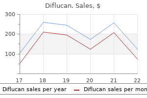
| Comparative prices of Diflucan | | # | Retailer | Average price | | 1 | DineEquity | 856 | | 2 | Ahold USA / Royal Ahold | 966 | | 3 | Williams-Sonoma | 842 | | 4 | Ross Stores | 197 | | 5 | Bed Bath & Beyond | 939 | | 6 | Publix | 862 | | 7 | Winn-Dixie Stores | 638 | | 8 | CVS Caremark | 190 | | 9 | OfficeMax | 431 |
Buy cheap diflucan 150 mgOsteophytosis of the vertebrae can be extreme and at times encroach on the spinal canal and intervertebral foram recognized to occur and should cause compressive myelopathy To conclude fungus zygomycosis diflucan 100 mg amex, plain radiographs are the primary imaging tools for the evaluation of suspected skeletal problems of metabolic and endocrine origin zarin anti fungal cream buy 200 mg diflucan fast delivery. The radiographic modifications are due to this fact predominantly diffuse or a minimum of multifocal involving many areas of the skeleton. An understating of the varied manifestations of those disorders including their findings on imaging help us reach an early diagnosis. Chapter 190 Skeletal Disorders of Metabolic and Endocrine Origin 3111 primary hyperparathyroid case with a number of brown tumors: a Orthopaedic Imaging: A practical approach, 4th edition. Technique and function in the preoperative analysis of primary with major hyperparathyroidism: 99m Company; 1990. Individual trabecular plates may be fractured or trabecular connectivity may be reduced. Thus, osteoporosis could be readily distinguished histologically from osteomalacia, during which mineralization of osteoid is impaired and the ratio of organic matrix to mineral is increased. The cumulative loss of bone mass ranges from 20% to 30% in men and 40% to 50% in ladies. Gastrointestinal Disease zz Alcohol-related liver illnesses zz Celiac disease zz Chronic energetic hepatitis zz Chronic cholestatic ailments zz Gastrectomy zz Inflammatory bowel disease zz Jejunoileal bypass zz Malabsorption syndromes zz Pancreatic insufficiency zz Parenteral diet zz Primary biliary cirrhosis zz Severe liver illness. It is often regulated by systemic and locally produced brokers and metabolic, dietary and mechanical elements. The regular balance between bone formation and resorption leads to maintenance of skeletal mass. In osteoporosis, the bone mass is decreased, indicating that the rate of bone resorption have to be more than that of bone formation. Skeletal progress is complete by the end of adolescence, however even after closure of the endochondral development plate, bone mass increases by radial development till peak bone mass is achieved at in regards to the age of 35 years. After a brief interval of balanced bone metabolism, bone resorption begins to exceed bone formation and skeletal mass decreases. The rate of bone loss in girls additionally Chapter 191 Osteoporosis 3113 Marrow-related Disorders zz Amyloidosis zz Hemochromatosis zz Hemophilia zz Leukemia zz Lymphoma zz Mastocytosis zz Multiple myeloma zz Pernicious anemia zz Sarcoidosis zz Sickle cell anemia zz Thalassemia. Miscellaneous Causes zz Ankylosing spondylitis zz Chronic obstructive pulmonary disease zz Congenital porphyria zz Epidermolysis bullosa zz Hemophilia zz Idiopathic hypercalciuria zz Idiopathic scoliosis zz Multiple sclerosis zz Rheumatoid arthritis zz Immobilization. Drugs-associated with Bone Loss zz Aluminum zz Anticoagulants zz Anticonvulsants zz Cigarette smoking zz Cytotoxic drugs zz Excessive alcohol zz Excessive vitamin A zz Glucocorticoids and adrenocorticotropin zz Gonadotropin-releasing hormone agonists zz Heparin zz Lithium zz Tamoxifen (in the premenopausal patient) zz Beta carotene zz Bile-acid binding resins. Primary Osteoporosis Type I (postmenopausal) osteoporosis: It occurs in girls between 51 and seventy five years of age with accelerated and disproportionate loss of trabecular bone. There is proportionate lack of both cortical and trabecular bone and the fracture sites are also those which comprise both kinds of bone, i. Juvenile Osteoporosis Juvenile osteoporosis is a rare illness which occurs in children between eight and 15 years. Clinical features embrace bone pain, fractures with minimal trauma (mostly metaphyseal fractures of distal tibia and vertebral crush fractures), lowered bone density at areas of latest bone progress and loss of peak. Biochemical investigations are regular and most forms of treatment are ineffective. There are numerous mechanisms answerable for this, and it has been shown by varied studies that both dose and length of steroid treatment correlate with bone loss. Hypogonadism: Whether due to primary gonadal failure or secondary to gonadotropin deficiency, hypogonadism is related to osteoporosis, particularly of trabecular bone. It is a nicely known fact that estrogen is necessary in stopping bone loss in regular premenopausal girls, and estrogen deficiency because of any cause can lead to osteoporosis. Both bone formation and resorption is increased in this dysfunction, but the formation is unable to match the resorption which leads to lowered bone mass. A primary lower in bone formation as a end result of lowered insulin motion has also been postulated. Hyperparathyroidism: Generalized loss of bone density is a prominent characteristic in major or secondary hyperparathyroidism which is adopted by endosteal resorption. Osteoporosis may be seen in patients with major hyperparathyroidism who may not have subperiosteal resorption or brown tumor or elevated serum alkaline phosphatase. Defective gene encoding resulting in decreased collagen formation of kind I results in osteoporosis. Osteoporosis also occurs in patients with homocystinuria as a end result of cystathionine synthetase deficiency, an autosomal recessive trait associated with ectopia lentis, numerous deformities of the extremities, psychological retardation, decreased pigmentation of hair and pores and skin and thromboembolism. The osteoporosis could also be as a result of the impact of homocystine or different metabolites in interfering with cross linking of collagen. It is characterized by arachnodactyly, posterior scalloping of vertebral bodies, excessive arched palate, lenticular subluxation and dissecting aneurysm. Gastrointestinal Diseases In diffuse liver illnesses, malabsorption or faulty metabolism of vitamin D may cause osteopenia. In addition many different gastrointestinal illnesses are related to osteoporosis. The neoplastic plasma cells secrete osteoclast activating factor which may be involved in mediating bone loss. Heparin or presumably different brokers released by mast cells or lymphoma cells will be the effector molecule for osteoporosis. Organ Transplantation Organ transplantation because the effective therapy for end-stage ailments of kidney, lung, liver and cardiac sufferers have improved, the variety of transplant sufferers have increased to an excellent extent on the planet. These patients require lifelong medicines, corresponding to steroids, immunosuppressant medicine and anticoagulation. Generalized skeletal ache is rare, and between fractures most patients are freed from signs. Pain often results from collapse of vertebra particularly in the dorsal and lumbar areas, sometimes acute and often radiates across the flank into the stomach. Type I osteogenesis imperfecta is an autosomal dominant form characterized by mildto-moderate bone fragility, blue sclerae and premature deafness. The advances in radiologic sciences have allowed the event of assorted noninvasive, correct and delicate techniques for determining the mineral content of bone. Standard Radiography Qualitative evaluation of bone mineral content from standard radiographs of skeleton is normally based mostly on subjective standards. The evaluation is commonly accomplished on metacarpal, calcaneum, talus, femoral neck, vertebral bodies, etc. Approximately greater than 30% bone loss should be there to be appreciated on plain radiographs. Subperiosteal and intracortical bone absorption, even delicate, may be appreciated on magnification radiography. Photodensitometry In this system, images of bone of interest and of reference aluminum wedge of known density are uncovered on the identical movie. The optical density of the bone is then compared with that of the wedge with photodensitometer. This provides data comparable to that of single photon absorptiometry but with barely decrease precision, sensitivity and accuracy.
150 mg diflucan with amexHowever fungus gnats damage cannabis buy diflucan 400 mg free shipping, contrast induced nephropathy has been related to gadolinium fungus gnats indoors 100 mg diflucan discount with visa, especially in sufferers with advanced renal illness. Further analysis can clarify the renal results of gadolinium in sufferers with renal insufficiency. Pruned, tortuous vessels with sluggish circulate with thinned renal cortex are demonstrated. Renal artery stenosis is most precisely recognized on angiography and may be treated by angioplasty. In a standard well-hydrated kidney, sharp corticomedullary differentiation is seen on T1W sequence because of excessive sign of cortex as compared to medulla. Loss of corticomedullary differentiation on T1W spin echo sequences is a sensitive indicator of parenchymal Radionulceotide Imaging Scintigraphy supplies imaging based mostly diagnostic info on renal construction and performance. Radionucleotide agents are excreted by kidneys; hence, they want to be judiciously employed in sufferers with renal failure. The most necessary risk issue is pre-existing renal insufficiency (serum creatinine >1. Adequate hydration, dose reduction and use of non-ionic contrast media are recommended as preventive measures. When obstruction is ruled out an analogous picture of enlarged easy kidneys with regular or effaced accumulating system is seen that signifies parenchymal disease of recent origin, which is probably reversible. As tubules are crammed with mobile infiltrates, contrast leaks into the interstitium by way of the broken basement membrane; hence, faint or nonopacification of pelvicalyceal system occurs. Other causes embrace extreme trauma with shock, sepsis, transfusion reaction, extreme dehydration, burns, peritonitis and toxins. Although large numbers of situations are related to acute cortical necrosis, the pathophysiology sadly stays unclear. For these surviving the early section, clean renal shrinkage happens over a number of months. Radiographically, distinctive tram like or egg shell calcification of cortex could also be seen which begin showing as early as 24 days after the onset of disease. Calcification of cortex causes dense cortical echoes with distal acoustic shadowing. The three diagnostic options are (a) enhancement of the medulla, (b) nonenhancement of the renal cortex, and (c) lack of excretion of distinction medium into the amassing system. Usually, markedly giant kidneys are seen; nevertheless, sometimes asymmetrical renal involvement or focal renal mass could additionally be seen. Imaging Features In leukemic infiltration of kidney, the nephrogram is faint and the collecting system is attenuated. Pelvicalyceal system is often filled with blood clots or uric acid stones which seem as filling defects. Amyloidosis Amyloidosis is a diverse group of illnesses which have extracellular deposition of an insoluble fibrillar proteinaceous substance with a beta sheath configuration. Amyloidosis may be categorized as follows:32 zz Primary Amyloidosis: Without pre-existing or coexisting disease. Imaging Features Bilateral renomegaly with diminished to regular opacification of amassing system is seen on urography. Enhanced uptake of Gallium citrate in the kidneys on renal scintigraphy can be reported. Calcification of renal papillae could happen in analgesic induced Chapter 108 Renal Parenchymal Disease and Renal Failure 1723 Heredofamilial amyloidosis associated with familial mediterranean fever. Older males are more affected than girls, 33 normally presenting with nonspecific symptoms like weight loss, fatigue and weak spot. Kidney is affected in 80% patients with secondary amyloidosis and 35�40% sufferers with main illness. Isolated involvement of the renal pelvis, ureter bladder, urethra, prostate seminal vesicle, and retroperitoneum can happen. Nausea, vomiting, anorexia, weight reduction and progressive weak point are the presenting features. Nephrocalcinosis resulting from hypercalcemia, uric acid calculi, renal infections and amyloidosis may further complicate the sickness. Imaging Features Urography shows enlarged clean kidneys with faint opacification indicating impaired renal operate. Injection of iodinated distinction media is hazardous in patients of multiple myeloma as it precipitates myeloma proteins within the renal tubules. Decrease in dimension occurs with development of disease course of, clean define is, nevertheless, maintained. Nephrogram is diminished with variable excretion into normal pelvicalyceal system. Rapid deterioration in renal function or sudden onset nephrotic syndrome indicates renal vein thrombosis, a complication of amyloidosis. Bilateral increased delayed uptake of Gallium 67 when other causes of irregular gallium activity are excluded. Patient presents with hemoglobinuria, iron deficiency anemia and venous thrombosis. Similar imaging features are seen in intravascular hemolysis like in malfunctioning prosthetic valves, sickle cell anemia, hereditary spherocytosis and thalassemia. Interesting radiological appearance of calcification or ossification in the amyloid deposits may be seen. Other causes for calcification of pelvicalyceal system are tuberculosis, leukoplakia major carcinoma of renal pelvis and renal calculus. Chronic Renal Parenchymal Disease In contrast to the capacity of kidney to regain back its perform following acute renal insult, renal injury of more prolonged nature usually results in progressive and irreversible lack of nephrons. Such discount in renal mass subsequently leads to bilaterally small easy kidneys. Radiological options, like those in acute renal parenchymal disease are overlapping in most of the causes of continual renal parenchymal illness. However, some conditions present attention-grabbing radiological appearances and are mentioned here. The various causes of bilateral small clean kidneys are listed in Flow chart 5, the circumstances resulting in morphologically small, unilateral Multiple Myeloma Multiple myeloma is a plasma cell dysfunction which originates in the bone marrow and is characterised by involvement of the skeleton at multiple websites. The neoplastic plasma cells produce excess immunoglobulins and Bence�Jones proteins are characteristically current within the urine. Note made of simple cortical renal cyst in left upper pole (white arrow) kidneys are additionally cited. Renal Papillary Necrosis Necroses of the renal papillae not only have many causes but in addition many radiological forms. Parenchymal diseases affecting the papillae and calyces are identified on urography. In mild circumstances the kidney dimension and performance are normal, and the abnormality is proscribed to the papillae only. Other causes are diabetes, sickle cell nephropathy, obstruction with infection, renal vein thrombosis, dehydration and extended hypotension. In early papillary necrosis ischemia occurs within the renal papillae because of compression of the medullary vessels by inflammatory adjustments in the interstitium.
Diflucan 200 mg buy discount lineInternational myeloma working group consensus statement and tips concerning the present position of imaging methods in the prognosis and monitoring of a number of Myeloma fungus photos 200 mg diflucan trusted. Magnetic resonance imaging in a number of myeloma: Diagnostic and medical implications antifungal lozenges diflucan 400 mg purchase. Correlation of measured myeloma cell mass with presenting clinical options, response to treatment, and survival. Myeloma management tips: a consensus report from the Scientific Advisors of the International Myeloma Foundation. Acute vertebral body compression fractures: discrimination between benign and malignant causes utilizing obvious diffusion coefficients. The use of a dedicated knee coil is mandatory for a excessive quality research as a end result of it improves the sign to noise ratio. Magnetic resonance imaging ought to cut back the variety of diagnostic arthroscopies and select the sufferers for therapy oriented arthroscopy. Magnetic resonance has supplanted nuclear scintigraphy in the characterization of osteonecrosis and can be utilized to assess the integrity of overlying articular cartilage surfaces. The smaller buildings, such as the posterolateral nook are also assessed better. Patient Positioning the affected person is positioned supine, feet first with the leg in full extension. The knee is positioned in 10�15� of external rotation to realign the anterior cruciate ligament parallel with the sagittal imaging airplane. This is often the position of the knee in the relaxed state and no effort at externally rotating the knee needs to be made in the majority of sufferers. The knee is positioned in a dedicated knee coil to produce a uniform sign intensity across the picture and foam pads are used to immobilize the knee inside the middle of the coil. Slice Thickness Four millimeter sections are used for axial, coronal and sagittal images of the knee. In the children, three mm slices enable optimum medial to lateral joint coverage within the sagittal airplane and anterior to posterior coverage within the coronal plane. Imaging Planes and Pulse Sequences Acquisition of images in three orthogonal planes is helpful in defining and characterizing abnormalities. Menisci and cruciate ligaments are finest evaluated on sagittal images with coronal views for secondary visualization and affirmation of pathology. The articular cartilage surfaces of medial and lateral compartments are assessed in both coronal and sagittal planes. The patellofemoral joint including patellar aspect and trochlear groove is greatest seen on axial pictures. Intravenous gadolinium is useful in assessment of inflammatory arthritides as it causes enhancement of the pannus. The biomechanics of patellar tracking can be assessed with kinematic approach with a cine loop display of acquired images. Tibial attachments to the meniscus are made by way of meniscofemoral, meniscotibial and coronary ligaments of the joint capsule. Microanatomy of the Menisci the menisci are predominantly made up of sort I collagen arranged into bundles. Most of the bundles course circumferentially parallel to the long axis of the meniscus. A smaller number of fibers are oriented in a radial trend functioning as stabilizing tie fibers. With axial loading of the joint, the circumferential orientation of a lot of the collagen bundles permits for meniscal deformation, the development of "hoop stresses" and a comparatively even distribution of the load across the joint surfaces. Up to one-third of the peripheral meniscus is vascularized and innervated (the "purple zone") whereas the remaining internal two-thirds or more is strictly fibrocatilagnious (the "white zone"). The higher margin is identified as the superior articular surface whereas the lower margin is called the inferior articular surface. Most horizontal tears are also not amenable to restore and the surgeon typically resects both the superior or inferior flap, leaving the other in situ. It sometimes has a striated look with some excessive sign within it especially at its insertion on the tibia. An irregular or wavy contour and disruption of fibers additionally suggests an entire tear. In partial tear, though the bulk of the ligament appears to be intact with a comparatively normal axis, there could additionally be localized angulation of the ligament on the site of fiber disruption. False-negative prognosis may end result from the formation of scar tissue with adherence of anterior cruciate ligament to the publish cruciate ligament simulating a standard course and signal of the anterior cruciate ligament. The invested deep fascia of the sartorius muscle, which overlies the gastrocnemius muscle, types the most superficial layer. The tibial collateral ligament, the primary assist construction, extends from the medial epicondylar area of the femur to the medial surface of the proximal tibia and spans about 8�9 cm in length. Magnetic Resonance Imaging Posteromedial corner accidents normally outcome from a valgus stress combined with rotational forces. It has a fusiform configuration and appears uniformly hypointense on all imaging sequences. A focus of marrow edema may be present when the posterior oblique ligament avulses. The arcuate ligament is as Y-shaped construction that represents thickening of the posterolateral joint capsule. These are seen as low sign intensity buildings on all pulse sequences and nearly uniform in thickness. Contour modifications, similar to thickening and irregularity, are extra typical of subacute or old accidents. Most complete ruptures involve the conjoined tendon and will produce a small avulsion of the styloid process of the fibular head. Complete tears of the popliteal tendon trigger enlargement of the muscle stomach and the retracted tendon terminates abruptly. Isolated popliteal injuries are rare, because most are related to concomitant accidents of the arcuate ligament complicated. The tendons of the gastrocnemius muscular tissues together with the soleus tendon type the Achilles tendon, which inserts on the posterior tubercle of the calcaneus. The major motion of the gastrocnemius muscle is plantar flexion of the foot but in addition serves as a passive supportive structure of the posterior joint capsule. The gastrocnemius muscle arises as two heads from the posterior surface of Magnetic Resonance Imaging Gastrocnemius accidents mostly are attributable to hyperextension of the knee or when the tibia posteriorly dislocates throughout knee flexion. Magnetic resonance imaging can assess the chronicity of the hematoma by the overall appearance of the products of hemoglobin degradation.
Syndromes - Problems with reasoning
- Brain damage caused by increased body temperature
- Rash
- Gargle several times a day with warm salt water (1/2 teaspoon of salt in 1 cup water).
- Small or dilated pupils (not reactive to light)
- Heparin
- Feeling weak or tired more often than usual, or with exercise
- Abnormal placement of the heart toward the right side of the chest instead of the left
- Certain high blood pressure drugs
Discount diflucan 50 mg with amexSex: In females fungus resistant fescue 150 mg diflucan cheap with visa, carcinoma breast is liable for majority of all bone metastases adopted by carcinomas of thyroid fungus how to get rid 50 mg diflucan generic free shipping, kidney and uterus. In males, carcinoma prostate is the commonest primary focus, followed by carcinoma lung. Blood borne metastases are unusual and often lytic, but might not often be osteoblastic or combined. Carcinoma of the uterine body is an adenocarcinoma and like that of the ovary, could produce osteoblastic metastases, particularly of the vertebrae. The irregular uptake on scintigraphy depends upon intact local blood flow and increased osteoblastic activity. Lesions which outgrow their blood supply or stimulate no osteoblastic response may seem as photopenic or "chilly spots" Occasionally a. Patchy rib lesions are also seen, but renal uptake is absent metastases than sclerotic metastases. Other features are the diaphyseal location, involvement of vertebral our bodies and then pedicles, the lack of bone enlargement, absence of florid periosteal response, tumor bone or gentle tissue mass. No one investigation, aside from biopsy, can provide a particular diagnosis of a metastasis or its web site of origin. Radiographic data mixed with information regarding the medical presentation, web site of the lesion, and the age of the patient permits formulation of a reasonable diagnosis typically. Metastatic seeding in the bone marrow is characterised by long T1 leisure times, whereas T2 relaxation instances are variable, depending on tumor morphology. Bone and soft tissue tumors in Magnetic Resonance Imaging in Orthopaedics and Sports Medicine, 3rd edn. Monitoring therapeutic response of major bone tumors by diffusion-weighted picture: initial outcomes. Ultrasound in musculoskeletal tumours with emphasis on its function in tumour observe up. Differential Diagnosis A solitary metastasis might resemble a primary malignant bone tumor, a primary benign tumor of aggressive nature. Post radiation sarcomas: a evaluate of the scientific and imaging options in sixty three circumstances. Sarcoma in Paget illness of bone: Clinical, radiologic, and pathologic features in 22 instances. Benign and malignant cartilage tumors of bone and joint: Their anatomic and theoretical foundation with an emphasis on radiology, pathology and clinical biology. Clear cell chondrosarcoma: Radiographic, computed tomographic and magnetic resonance findings in 34 sufferers with pathologic correlation. Fibrosarcoma and malignant fibrous histiocytoma of lengthy bone: Radiographic options and grading. A full rupture of the gastrocnemius head is related to retraction of the muscle stomach. Knee dislocations are essential as a result of they produce extensive disruption of the ligaments that stabilize the knee and the encircling gentle tissue structures, together with the popliteal artery. Complete disruption of 1 or each of the collateral ligaments is expected with this severe injury. Frequently, the sample of bone contusions permits definition of the type of knee dislocation. This is a vital observation as a end result of posterior and posterolateral dislocations have a high association with peroneal nerve harm. Magnetic Resonance Imaging A knee dislocation is classed according to the place of the tibia relative to the femur. There are 5 different varieties of dislocations: (i) anterior, (ii) posterior, (iii) lateral, (iv) medial, and (v) posterolateral. Segond Fracture the Segond fracture, or avulsion of the anterolateral aspect of the lateral tibial plateau, is a radiographic entity that signifies underlying trauma to the anterolateral nook constructions of the knee. This injury is seen most often in people who take part in long-distance running and cycling. The layered configuration of the quadriceps tendon enables discrimination between partial and full tears. Discontinuity of any of the tendinous layers is according to partial tears usually involving the rectus femoris element. Transection of all the layers is diagnostic of a complete rupture and could also be associated with a mass of edematous tissue and hemorrhage seen as excessive sign on T2W pictures. The patellar tendon also has homogeneous low sign depth look aside from small occasional triangular areas of intermediate sign depth instantly under the patella and adjacent to the tibial tuberosity. A massive collection of adipose tissue, the infrapatellar fats pad of Hoffa, rests simply posterior to the patellar tendon. In symptomatic patients, the findings may be accompanied by patellar tracking disorders in as many as 45% of patients. Acute Patellar Tendon Disruption A minority of patellar ligament injuries are complete tendon disruption ensuing from acute macrotrauma within the setting of sports-related activity. Femoral Trochlear Groove the femoral trochlear groove offers a mechanical restraint that helps stabilize and function a information for the patella as it articulates with the groove throughout joint function. In the axial aircraft the posterior apex of a traditional patella is centered directly above the intercondylar femoral sulcus. Shallow sulcus angles (larger sulcus angle measurements) predispose to patellar instability. Affected sufferers are both athletes and nonathletes who expertise persistent anterior knee ache that localizes to the inferior patellar pole on bodily examination. Most affected person show patellar alignment abnormality, such as patella alta or lateral subluxation. Patients typically reply well to conservative remedy, which usually entails taping the superior pole of the patella. The avulsed free end of the retinaculum is commonly frayed and thickened and fluid could dissect around a muscular slip of the vastus medialis muscle. A low using patella (patella baja or profunda) occurs in quadriceps tendon rupture but can also be found with paralytic neuromuscular issues, achondroplasia and juvenile rheumatoid arthritis. Patellar shape: Wiberg has proposed a 3 part classification to embody the vast majority of patellar aspect configurations. This procedure should be repeated at three different part places so that the entire tour of the patella could be evaluated as it articulates with the femoral trochlear groove. Compared with the incremental, passive positioning technique, a more physiological examination is obtained and in certain instances abnormal patellar tracking is extra obvious during energetic motion. Motion Triggered Cine Technique In this method, a particular nonferromagnetic gadget incorporates a trigger system that senses the movement of the patella. The affected person is positioned supine on the positioning system and a single patellofemoral joint is flexed and prolonged repeatedly while gradient echo photographs are obtained using a circular surface coil. Articular cartilage function is lost with injury to chondrocytes, collagen or proteoglycans. With more severe damage cracks, fissures, and focal defects form within the cartilage. Because of its hydropic composition normal hyaline cartilage demonstrates intermediate signal intensity on T1-weighted pictures compared with the low sign intensity of the cortex and fibrocartilaginous menisci.
50 mg diflucan cheap otcSagittal sonogram demonstrating the echogenic line (arrowhead) that extends from the renal sinus to the perinephric fats hilum antifungal body wash cvs purchase 200 mg diflucan otc. Accessory renal arteries may come up from the aorta in as many as 20% people fungus yard pictures buy diflucan 400 mg low cost, either superior or inferior to the main renal artery. The renal arteries usually divide into anterior and posterior divisions that lie anterior and posterior to the renal pelvis, respectively. These divisions give rise to the segmental arteries which branch further throughout the renal sinus, forming interlobar arteries that penetrate the renal parenchyma. These terminate in arcuate arteries that curve around the corticomedullary junction giving rise to cortical branches. Occasionally it might be difficult to differentiate a small avascular tumour from a hypertrophied column of Bertini when further investigations perhaps required. It was thought to represent connective tissue on the junction of the event of anterior and posterior components of the kidney. However, subsequent reassessment means that this line represents an extension into the parenchyma of hilar/sinus fat in sufferers with a deep renal sinus, rather than a real airplane of fusion between embryological elements. An enhance in fat content of the renal sinus can occur in obese individuals, in renal sinus lipomatosis, and in cases of parenchymal atrophy. A mild distension of the amassing system can occur due to physiological filling. This can be seen in a fluid loaded topic,a patient on diuretics, diabetics, restoration part of acute tubular necrosis, single kidney, neonatal kidney and patients with an overdistended bladder. The proper and left posterior oblique positions can also be used to identify the vessels in the midline. Throughout the course of examination of renal vessels, colour Doppler is regularly switched on to verify the character and course of circulate. The optimum pulse repetition frequency is chosen to detect moderate flow velocities, though it could have to be modified to detect high velocities if a stenosis with aliasing of colour signals is current. With the system set to detect low or reasonable flow velocities, circulate could be recognized in almost all sufferers in the vessels at the renal hilum. Angling of the probe medially from the best or left flank will enable assessment of the intrarenal vessels. Though the hilar and interlobar vessels are demonstrated in all patients, the arcuate and striate arteries could also be seen solely in slimmer sufferers. The use of ultrasound contrast material can additional improve visualization of parenchymal vessels. There is a speedy systolic upstroke, which is occasionally followed by a secondary slower rise to peak systole. Further, Doppler Evaluation the renal arteries arise from the aorta, barely beneath the origin of the superior mesenteric artery. Ureters the ureter is an extended (30�34 cm) mucosal lined tube varying in diameter from 2�8 mm. The proximal ureters are finest visualised in a coronal indirect view, using the kidney as a window. An attempt may be made to observe the ureter upto the bladder using the identical method. The normal renal artery spectral waveform exhibits a steep systolic peak and a smooth downslope to diastole; (B) Spectral waveform in an intrarenal vessel exhibiting decrease velocities Chapter ninety four Ultrasound of Urogenital Tract: Techniques and Normal Appearances 1501 (transrectal/transurethral) can give wonderful element of the bladder wall. The full bladder and postmicturition residual volume may be calculated using the formulation for a prolate ellipsoid (0. Common indications for sonography of the lower urinary tract are as follows: z Determination of the existence and price of urine flow by way of the vesicoureteric junction in patients with dilated ureters z Determination of pre- and post-void bladder volume z Detection of bladder calculi or mass z Detection and quantification of bladder wall thickening z Guidance for diagnostic or therapeutic interventional procedures Transrectal and transurethral scanning can consider the layers of the bladder wall and consider the extent of a mass lesion through tissue planes in and around the urinary bladder. The normal penile urethra is seen as an anechoic tubular construction when distended. The feminine urethra may be visualized on transabdominal scanning in 35% of the sufferers and in 100% of the patients with a catheter in place. It can also be well recognized in all patients by translabial or endovaginal scanning with the probe placed at the introitus or simply partially inserted into the vagina. Normal Sonographic Appearance the conventional distended bladder is an anechoic construction occupying the midline of the true pelvis. It has thin walls (less than 3 mm within the distended state and 5�6 mm when nondistended), that are regular and clean. Transabdominal evaluation of the ureteral jets is helpful to assess for any proximal obstruction. On grey scale, a stream of low level echoes could be seen coming into the bladder from the ureteric orifice. Density variations between the jet and bladder urine allow its sonographic visualization. Depending on the state of hydration, the jet frequency may vary from lower than 1 per minute to continuous flow; however, each side must be symmetrical in a wholesome particular person. Detection of these jets excludes full ureteric obstruction and establishes renal perform. The prostate is visualized posterior to the bladder in both axial and sagittal planes. The mostly used commercially out there probes hearth from the top and can be utilized for both transrectal and transvaginal imaging. The patient is examined in the left lateral decubitus place with the knees flexed. In the sagittal aircraft, the gland is systematically surveyed from right to midline to left; whereas within the axial airplane, cranial to caudal viewing is completed from a plane just superior to the prostate (showing the seminal vesicles) to the apex of the prostate caudally. The volume measured can be converted to weight as a outcome of the specific gravity of prostatic tissue is 1, thus 1 mL of prostate tissue is the identical as 1 gram. The presence of anal or rectal strictures or advanced rectal carcinoma could disallow the transrectal technique. Common Indications for sonograhic analysis of the prostate are: z To quantify prostate quantity z Assessment of a palpable nodule z Evaluation of infertile patients z Guided prostatic biopsy. It measures approximately 4 cm in length (cephalocaudal), 4 cm in transverse diameter and 3 cm in peak (anteroposterior); the conventional volume (weight) ranges from 20�25 mL (g). The border of the prostate appears sharply defined, except on the posterolateral margins the place the neurovascular structure enters the prostate gland. The central zone surrounds the ejaculatory ducts and is the location of the primary lesion in less than 10% of circumstances. At the bottom of the gland, the seminal vesicles immediately adjoin the central and peripheral zones. In the midsagittal section the collapsed urethra is seen as an echogenic line surrounded by the hypoechoic easy muscle forming the internal urethral sphincter. With colour Doppler, significantly utilizing the ability mode, the prostate is seen as a very vascular structure. The capsular and urethral arteries are simply seen, and branches to the inside gland and peripheral zone are normally very distinguished. They are imaged in the long axis on the axial images and in cross-section on sagittal pictures. They are up to 1 cm in width, however often they could be very large in regular males.
Buy 100 mg diflucan with mastercardPseudoaneurysm Pseudoaneurysms happen secondary to trauma or infection and consist of leakage of blood into the confined perivascular area on the site of a vessel wall disruption antifungal for face discount 50 mg diflucan. In large-neck pseudoaneurysms antifungal essential oil blend 200 mg diflucan generic with amex, a stent placement mixed with coil embolization has been described. An important prerequisite for chemoinfusion/chemoembolization is the presence of a patent portal vein with hepatopetal flow. The bilirubin level should be lower than 3 mg/dL to perform chemoinfusion/chemoembolization safely. Initially, a superior mesenteric arteriogram is normally obtained to reveal a variant origin of hepatic artery (accessory or changed, originating from the superior mesenteric artery) and to reveal patency of the portal vein. Then, the celiac trunk and, subsequently, the widespread hepatic artery are catheterized and studied to outline the vascular anatomy. The involved lobar hepatic artery or, extra generally, the first- or second-order branches of this artery is subsequently catheterized by utilizing a microcatheter and the chemoinfusion materials is injected beneath fluoroscopic guidance. The tip of the catheter should be positioned distal to the cystic and gastroduodenal arteries. The most commonly used chemoinfusion mixture consists of 10 mL of iopamidol (Isovue), 20 mL of Ethiodol, and 60 mg of doxorubicin. The chemoinfusion is normally followed by embolization with slurry of gelatin sponge powder (Gelfoam). Lidocaine is intra-arterially administered to scale back pain after the chemoinfusion/chemoembolization treatment. Renal embolization is an various alternative to surgical removal of the kidney, and indications include end-stage renal disease or renovascular hypertension requiring unilateral or bilateral nephrectomy and renal transplant with native kidneys in situ. The procedure requires selective catheterization of the renal artery with additional advancement of the catheter so that the catheter is wedged or with the usage of a balloon occlusion catheter to decrease the potential of embolic materials spillage into the aorta. Postinfarction syndrome is comparatively widespread and characterised by pain, which could be managed with narcotics. Balloon dilatation could be carried out safely even in babies and may permit access to peripheral stenoses. Larger balloons up to 20 mm in size are used to deal with recurrent coarctation or peripheral pulmonary stenosis. High strain balloons (up to 17 atm burst pressure) are available with smaller sizes for fibrous stenosis or restenotic lesions. Embolotherapy is performed with superselective catheterization/embolization of the splenic artery through the use of embolic particles whereas the tip of the catheter is past the caudal pancreatic artery. Careful fluoroscopic control of the splenic area is required to restrict the total infarction to approximately 60% of the spleen. Other causes embody fibromuscular dysplasia (28%), atherosclerosis (8%), polyarteritis nodosa (2. The complexity of pathological adjustments within the wall of the aorta and widespread nature of involvement make surgical revascularization a really troublesome possibility. Due to these causes, nonsurgical revascularization techniques have been increasingly used in the remedy of this group of sufferers. Antihypertensive medication is stopped 24 hours earlier than angioplasty, aside from sublingual administration of 5�10 mg zz zz nifedipine if the blood stress is more than 170/110 mm Hg. The patients are treated with aspirin (175�330 mg) every day for three days earlier than angioplasty, and this therapy is sustained for 6 months after therapy. Blood stress medication is withheld for 24 hours after the procedure, apart from sublingual administration of nifedipine (5�10 mg) if the blood pressure is above 160/100 mm Hg. The diseased renal artery is selectively catheterized via another arterial sheath in the reverse groin and transstenotic strain gradient is measured. The angiographic catheter is changed by a commercially obtainable, appropriate sized balloon catheter by utilizing commonplace trade method. The diameter of the involved vessel is measured and a balloon catheter of identical size is used for angioplasty. Three to 5 inflations, for as a lot as 45 seconds each, are carried out till the balloon "waist" is now not current or has decreased substantially. Immediately after the procedure, transstenotic pressure is measured and an angiogram is obtained to assess the adequacy of angioplasty. Alternatively, the process can be accomplished via a single groin approach too. Theleftstenosiswas then subjected to angioplasty with a similar result 1964 Section 5 Pediatric Imaging configuration can also be used) are then used to dilate the lesion. The advantage of this approach is the avoidance of a second puncture, though the worth of hardware will increase. Pretreatment with ticlopidine (250 mg twice daily) beginning three days earlier than angioplasty is then advisable. A preshaped renal guiding catheter is positioned at the ostium of the diseased renal artery over an change guidewire positioned in a safe distal location within the artery. The number of the diameter and size of the stent is predicated on the angiographic morphology of the concerned artery. It is advisable to give sublingual nifedipine (5�10 mg) or an intra-arterial bolus of trinitroglycerine (100�200 mg) within the renal artery before stent placement. The stent is positioned throughout the lesion and released by inflating the balloon on the desired inflation stress for up to 30 seconds. A check angiogram is obtained at the end of the process to assess the adequacy of stent release. Intravascular ultrasound is a useful method to define the endpoint of intervention. Angioplasty is taken into account technically profitable if: (1) the aortic or renal artery lumen after angioplasty has lower than 30% residual stenosis (2) the arterial lumen is a minimum of 50% bigger than its pretreatment diameter, and (3) the stress gradient is lower than 20 mm Hg and has decreased no less than 15 mm Hg from the pretreatment gradient. Follow-up angiograms are carried out in sufferers with recurrence of hypertension, in whom contralateral nephrectomy of poorly or nonfunctioning kidney for residual hypertension is deliberate and in those sufferers who consent for the process. Stents have been occasionally used as a "bail-out" measure in salvaging an obstructive dissection in such conditions and rarely electively within the therapy of native stenosis. Stents present a direct aid of symptomatic obstructive dissection and are also helpful in the remedy of recurrent stenosis after profitable angioplasty. Until lately, it was felt that this diesease is characterized by skip areas of involvement. The findings of recent studies, using cross-sectional imaging strategies, recommend that nonspecific aortitis involves a continuous length of the aorta, producing mural and luminal modifications in some areas, and only mural adjustments within the intervening segments. The outcomes of cross-sectional imaging counsel that there are intensive wall modifications even in angiographically normal areas. In this regard, intravascular ultrasound could also be helpful in guiding the interventional procedures. Late reworking occurs in most patients and is liable for delayed scientific benefit regardless of poor technical success in some sufferers. Peripheral pulmonary artery stenosis can sometimes be handled effectively however at reasonable risk.
150 mg diflucan buy with amexThe hemorrhages are most often seen within the ends of lengthy bones fungus under nose 100 mg diflucan cheap visa, corresponding to femur antifungal ear drops purchase 150 mg diflucan mastercard, tibia, humerus. Upon healing, the subperiosteal hemorrhages calcify and forged shadows of elevated density within the soft the above findings constitute the basic signs of childish scurvy. These modifications are much less apparent as the affected person Clinical Findings presents with progressive irritability, with tender, edematous adults, being seen in patients affected by persistent extreme malnutrition. Biochemical Findings Pathophysiology the pathological modifications seen in scurvy are the end result of despair of regular mobile activity. At the growth plate, although cartilage proliferation is decreased, mineralization is unimpaired, ensuing within the zone of provisional calcification becoming extensive and dense. Thus, the trabecular bone mass is decreased in the zone of primary and secondary spongiosa. This is seen radiographically as a transverse band of radiolucency adjoining to the zone of provisional calcification. This zone, called Tr�mmerfeld zone represents an area of weakened bone and reveals an inclination to fracture. The zone of provisional calcification extends past the margins of the metaphysis, resulting in periosteal elevation and marginal spur formation. On healing, all adjustments are reversible on vitamin C remedy although a single progress arrest line could remain within the deposition per se. A generalized improve in bone density is seen which is due to osteoclastic response to the fluorine somewhat than fluorine Radiographical Findings 1. Osteosclerosis involves all bones however is most marked in the a thickening of the bony trabecular sample which then progresses to a dense, uniform, symmetrical sclerosis that obliterates the bony architectural landmarks. The cranium and tubular bones of the appendicular skeleton are rela tively spared within the sclerotic course of. The solely change in the calvarium may be a sclerosis of the base of the skull and posterior clinoids. Thickening of the cortex of the affected bones happen on the expense of the medullary cavity. Ligamentous calcification and ossification; significantly of sacrospinous and sacrotuberous ligaments is one other attribute radiographic feature. Neutron Activation Analysis In this method, neutrons from an accelerator or reactor bombard a small fraction of the total 48Ca within the body, altering it to 49Ca which is a radioactive isotope. Practically, the intensities of high-energy and low-energy photons are analyzed individually after the protons have handed via bones and delicate tissue. With use of a specific computing algorithm, the attenuation values of sentimental tissues are subtracted, leaving only the attenuation values of bone. Fan-beam scanners use wider beams (as in comparability with beforehand used pencil width X-ray beam) that allow extra speedy scanning (approximately 3�5 minutes per site; times are related for wholebody scans), improved picture high quality, and a spatial resolution of 0. It is based on measurement of scattered radiation from a source of 100�700 keV gamma rays and is used for calcaneum, backbone and radius. The precision of this methodology is about 3�5% and radiation dose varies from 200 to 2000 mrem. Usually the radius is measured at two sites, the mid diaphysis which consists of nearly 100% cortical bone, and the distal radial metaphysis which incorporates about 75% cortical bone and 25% trabecular bone. A major clinical limitation of this method, subsequently, is that it displays the standing of peripheral lengthy bone and measures primarily the cortex. The chief benefit is that the gamma ray power of the source is larger (153Gd with photons of forty two and 100 keV), and it has a uniform path length. This method yields integral of all mineral inside the scan path together with the vertebral our bodies, endplates and posterior parts. The major drawback is that vertebral compression fractures with callus formation, kyphoscoliosis, articular aspect hypertrophy, diskogenic sclerosis, marginal osteophytosis and extraosseous calcification (aorta) are also included within the integral measurement and should result in inaccurate and poorly reproducible vertebral measurements. In healthy subjects, the precision is in the order of 2�3% with accuracy of 4�10% of coefficient of variance. Nor can it help those due entirely to elevated bone density (within a fixed volume of bone). Quantitative readings are obtained from a area of interest over trabecular bone encompassing 3�4 cm3 of each vertebral body and from four completely different reference options within the phantom. The precision of this technique is 1�3% for single power (80 kVp) and 3�5% for dual energy (80 kVp/140 kVp) strategies with accuracy of 5�10%. In a examine of 55 patients of prostate cancer who underwent orchidectomy the creator discovered that a statistical discount in vertebral trabecular bone mineral density was observed in all sufferers within six months of orchidectomy. The creator found a significant lower in trabecular bone mineral density at all vertebral levels with a median decrease of 26. Accurate and constant positioning of the forearm and reference strains are important in any longitudinal or multicenter research for comparable results. Quantitative Ultrasound Ultrasound has been efficiently used for a number of years for nondestructive materials testing. Both scattering traits and ultrasound velocity modifications have been used to consider mechanical competence and detect the presence of injury in both supplies and buildings. In addition to assessing bone density these variables reflect parameters, corresponding to elasticity and microarchitecture that are necessary in assessing fracture threat in osteoporotic topics. The ultrasound wave is produced in the form of a sinusoid impulse by special piezoelectric probes, and is detected once it has handed by way of the medium; there are two distinct probes, emitting and receiving, and the skeletal phase for analysis is positioned between them. Numerous efforts have been made to develop trabecular bone imaging of peripheral websites, such because the radius, tibia and calcaneus. Recently clinically useful pictures of femur (the deeper structure) are obtained by Krug et al. Secondly, the gradient-echo has to be acquired instantly after the excitation pulse in order to keep away from sign loss because of T2 relaxation. The easiest sequence that fulfills the aforementioned necessities is a primary gradientecho sequence. Image postprocessing: There are many various approaches for extracting structural info from 3D pictures of the High-resolution Bone Imaging the importance of assessing the microarchitectural make-up of addition to its mineral density within the context of osteoporosis has been emphasised in a quantity of publications. Information regarding structure, topology, and orientation of the trabecular bone network as well as cortical thickness and area can be extracted from the photographs by making use of digital postprocessing techniques. Trabecular bone analysis: Structural parameters are commonly divided into three courses, together with scale, topology and orientation. Topology may be assessed by investigating the plate- or rode-like structure of the community. And lastly orientation strategies characterize the degree of anisotropy of the structure. Image acquisition scan set-up: A normal, manufacturer really helpful scan protocol has been used within the majority of the printed literature to date.
Effective diflucan 50 mgIn 30% antifungal washing detergent 200 mg diflucan generic visa, the appearance is of a multilocular cystic mass with intervening solid areas fungus under fingernails discount diflucan 400 mg without prescription, which may calcify. Doppler An abnormal Doppler sign can be seen within the ipsilateral renal artery as an increase in velocity, spectral broadening and turbulence. Intraoperative ultrasound is being used in nephron sparing surgical procedure to delineate actual extent of the mass and venous thrombi. Computed Tomography Ultrasound Renal adenocarcinoma is detected as a nicely encapsulated stable mass. Clear cell carcinomas enhance to a greater degree than different subtypes of malignant lesions, especially papillary carcinoma. The criterion of 1 cm as a threshold has proved to be unsuccessful in differentiating reactive nodes from metastatic adenopathy. Based on T1-weighted pictures, a 15�20% enhancement on scans 3�5 minutes after gadolinium is considered diagnostic. Subtraction (gadoliniumenhanced fat-suppressed T1-weighted images�unenhanced fat-suppressed T1-weighted image) is a simple, reliable technique. Enhancing papillary projections at periphery of a cystic hemorrhagic mass are seen. Angioinfarction has a limited function, in massive tumors and as palliative procedure to control pain, hematuria in inoperable/ unfit for surgical procedure sufferers. False-positive results have also been seen in sufferers with benign inflammatory means of the kidney or benign tumors. Flurodeoxyglucose-positron emission tomography may have a role in evaluating distant metastasis and in the differentiation between recurrence and posttreatment modifications. Staging of Renal Cell Carcinoma7-11 Staging is a crucial part of analysis of a affected person with renal mass. Some signs like stranding of perinephric fats fascial thickening, obliteration/blurring of fats, adrenal involvement, seen collaterals are thought of suggestive for extracapsular unfold. Perinephric stranding might end result from edema, congestion and obliteration of fats may be as a outcome of mass effect. If tumor extends only into subdiaphragmatic cava, then a flank strategy is sufficient. On the best aspect it might be difficult to distinguish between intraluminal tumor thrombus and extrinsic caval compression brought on by a large primary tumor or enlarged lymph nodes. Falsepositive rates of 58% have been reported, nevertheless, when a size criterion of 1 cm is used, owing to reactive or different benign nodal disease. These false-positives are more frequent in patients with tumor involvement of the renal vein and tumor necrosis. A normal node will take up this particle and create a drop in T2 signal, whereas no drop in sign is seen in metastatic nodes. Bone scan is carried out solely in patients with bone ache or raised alkaline phosphatase. Bone scan could also be negative because the lytic metastasis produce little osteoblastic response. Follow-up Most recurrences happen inside three years with a median relapse time of 1�2 years. Normal constructions might migrate into the renal fossa after nephrectomy and may simulate recurrent tumor. The liver, ascending colon, 2nd a part of duodenum, pancreatic head and small bowel may migrate into the right side. Flurodeoxyglucose-positron emission tomography may also play a task in comply with up with a high constructive predictive worth. Renal sparing surgical procedure is now becoming a broadly accepted technique for removing small cancers. These patients have a decreased risk of continual renal insufficiency and proteinuria. The central tumors are more difficult to ablate than the peripheral exophytic ones. It has been suggested that stage somewhat than tumor grade is the main prognostic issue for urothelial tumor of the upper tract. There may be obstruction of kidney with non opacification of the accumulating system (phantom calyx) due to extensive areas of parenchymal infiltration. Calices may be amputated or obliterated because of malignant infundibular structuring. Retrograde or percutaneous antegrade pyelography may be useful in such conditions to delineate the location of obstruction and to obtain brush biopsy. Other non particular findings embody hydronephrosis, pyelocalyceal wall thickening. The central mass expands the kidney symmetrically and with centrifugal extension preserves the form. Involvement of renal parenchyma could additionally be seen as a hypoenhancing mass involving parenchyma or heterogeneous irregular hypoenhancement disrupting the normal parenchyma. A different appearance is thickening of collecting system, urothelium or ureteral wall. Squamous Cell Carcinoma Squamous cell carcinoma of the renal pelvis is comparatively rare tumor. It usually entails the renal parenchyma and perinephric tissue and may present with metastasis. Intravenous urography could reveal nonvisualization, hydronephrosis, a central mass or a pelvicalyceal filling defect seen in 83% of sufferers. Renal medullary carcinoma is similar and arises from calyceal epithelium, in or close to the renal papilla, from which it grows in an infiltrative pattern. It is seen in young African-American youngsters and adults, seen exclusively with sufferers of sickle cell trait and generally has metastasis at time of presentation. They are commonest in sufferers greater than forty years of age and they current with hematuria, stomach distention, weight loss or pain. Leiomyosarcoma is the most common type of renal sarcoma, comprising half of all renal sarcoma. Liposarcoma of the kidney arises from the renal capsule and appears as a big retroperitoneal mass with macroscopic fat. Extranodal unfold of lymphoma often affects the genitourinary system with the kidneys, being most commonly concerned organ. Renal involvement is usually asymptomatic, detection normally occurs at imaging research. Despite the high prevalence, imaging studies demonstrate involvement in only 3�8% of patients. Renal lymphoma has a big selection of imaging appearances depending on the pattern of tumor proliferation. Malignant lymphocytes attain the renal parenchyma by the use of hematogenous spread and proliferate within the interstitium, using nephrons, tubules and blood vessels as scaffolding for further development. If it follows this infiltrative pattern, kidneys enlarge, maintaining the reniform shape. Some tumors spread by the use of contiguous extension from the retroperitoneal illness, penetrating the renal capsule.
|
